Archives

What a maggot’s genes can reveal about a corpse

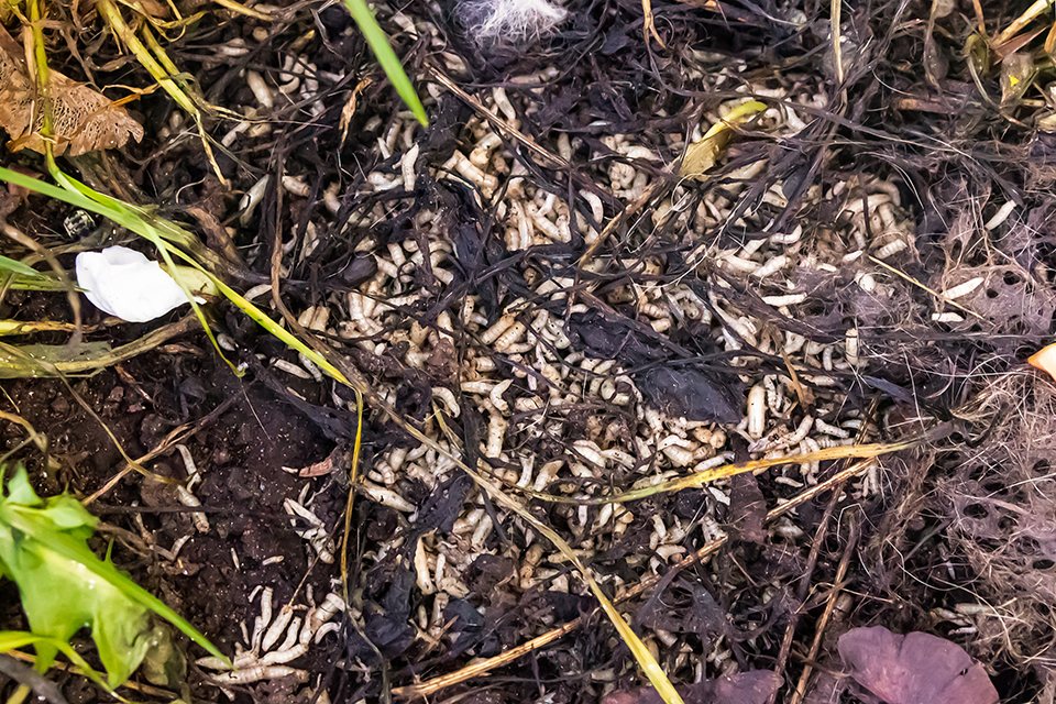
At a crime scene, a single fly larva can sometimes reveal more about the time of death than an autopsy. For decades, insects have been among the most reliable indicators for dating a death. When a body is found several days or several weeks after death, the classic methods of forensic medicine lose precision and forensic entomology takes over. Blowflies and other necrophagous insects lay their eggs on a corpse on a fairly regular schedule, one that can be worked backwards. By measuring the size of the larvae and identifying their developmental stage, the expert estimates their age and, allowing for the temperature at the scene, infers the minimum time since death [3].

This method remains the foundation of the discipline, but it soon reaches its limits. Beyond a certain point, larvae stop growing in any visible way while continuing to develop inside. At the same size, 2 larvae may in fact be several hours apart, depending on temperature or on what they have eaten. At the final larval instar in particular, size alone can no longer settle the matter [2].

Picture a body found in a wood a few days after a missing-person report. Investigators collect the largest larvae, the ones that have reached the final instar. Comparing their size against the growth curves for the species, adjusted to the temperature recorded on site, they arrive at an approximate time of egg-laying, with a margin of around 24 hours. That margin can change everything, because it decides whether the flies laid their eggs before or after a suspect passed through. Since the larvae hardly grow any further at this stage, their size cannot reduce the uncertainty, and reading their genetic activity could help to close that gap.

When appearance is no longer enough, look to the genes

Faced with this difficulty, a Chinese team led by Jiangtao Mei has taken stock, in the journal Legal Medicine, of a promising line of research [1]. That line, transcriptomics, means looking not at the DNA itself but at the genes actually switched on in the cells at a given moment. A person’s DNA does not change over a lifetime, and its sequence is fixed. The genes it uses at any given moment, by contrast, shift with the stage of development. Observing that activity is a little like photographing the biological state of the larva at the moment it is collected.

The idea goes back some years. As early as 2011, Tarone and Foran showed that the age of green bottle fly larvae, Lucilia sericata, could be estimated more precisely by adding, to measurements of size and stage, the activity level of a few well-chosen genes. The gain was clearest exactly where size fails, at the most advanced stages [2]. In the pupae of Calliphora vicina, later work identified genes whose activity changes at each step of development, making it possible to build targeted assays for dating those pupae [5]. The value of the review by Mei and colleagues is to bring these scattered studies together and give them coherence. Across several species of forensic interest, the activity of certain genes follows a course regular enough to serve as a useful complement to estimation by eye [1].

The choice of these genes is anything but arbitrary. They are, above all, genes tied to processes that unfold in a regular way, such as the cell’s production of energy or the hormonal signals that govern each moult. Metamorphosis, for instance, is triggered by a hormone, ecdysone, which switches on a whole cascade of genes as development proceeds. Because they follow a well-regulated biological programme, these genes offer the most reliable markers [5].

A shift seen elsewhere in the forensic sciences

This move from the visible to the molecular reaches beyond entomology. In human forensic genetics, a DNA profile is no longer the whole of identification. Other molecules are now being asked what they can reveal. The RNA in a biological trace helps to establish which tissue or body fluid it came from, while the methylation marks fixed on the DNA give an indication of the age of the person who left it. Forensic anthropology is undergoing the same change, with molecular analysis coming to support the traditional examination of bone. Entomology is taking the same turn. Here too, outward observation does not tell the whole story, and the aim is to understand what is happening in the insect’s cells at the moment of collection.

A recent study gives a good picture of this approach. Unable to work on human bodies, one team placed 3 pig carcasses in the open air and tracked, at the same time, the succession of insects, the microbial populations and the degradation of RNA in the muscle [4]. Each of these lines of evidence covers a different window. The insects colonised the carcasses within hours and were still present more than 40 days later. Muscle RNA degraded at a still-readable rate until about 240 hours after death, roughly 10 days. The microorganisms, for their part, dated decomposition to within 3 or 4 days [4]. Cross-referencing these sources rather than relying on any single one sharpens the estimate, and Mei’s review regards this combined approach as the most promising [1].

Findings still far from the courtroom

The authors nonetheless call for caution [1]. Most studies cover only a few species, reared in the laboratory under stable conditions far removed from a real death scene. Yet gene activity depends on many things, first among them temperature, but also the larvae’s food, the stress they undergo and the genetic differences between populations of the same species from one region to another. A gene that behaves regularly in an incubator at a fixed temperature will not necessarily respond the same way on a body left outdoors, where the temperature varies and several insect species compete over the same corpse.

There is also the question of cost and time. Analysing gene activity calls for far more complex equipment and expertise than measuring a larva against a ruler, and the results take longer to interpret. No one, then, is proposing to abandon the current methods, which rest on temperature-linked development curves validated by decades of field data. The strongest constraint remains the criminal trial. For evidence to stand up in court, a method must have demonstrated its reliability, its reproducibility and a known error rate under conditions close to those of the field, which is not yet the case for these markers [1]. The real challenge, for the authors, is therefore to pinpoint the specific cases where genetic analysis would add something, for example when a larva’s stage is too ambiguous to be settled by eye, or when the temperature at the scene could not be measured properly [1].

A discipline still reinventing itself

Forensic entomology was long seen as a field science, a matter of observation and experience. Recent work on gene activity shows that it is in fact following the same path as forensic genetics and forensic anthropology, which have themselves moved to molecular tools. It is not yet known when these markers will be reliable enough for routine casework. A larva, though, is more than its length. Its gene activity carries its own record of the time since death.

References

[1] Mei J., Liu S., Tao H., Xia S., Wang Y. (2026). Transcriptomics in forensic entomology, research progress and prospects. Legal Medicine, 81, 102801.

[2] Tarone A.M., Foran D.R. (2011). Gene expression during blow fly development, improving the precision of age estimates in forensic entomology. Journal of Forensic Sciences, 56 (suppl. 1), S112-S122.

[3] Pigoli D. et al. (2023). Estimation of temperature-dependent growth profiles for the assessment of time of hatching in forensic entomology. Journal of the Royal Statistical Society, Series C (Applied Statistics), 72 (2), 231-253.

[4] Wang Y. et al. (2021). Dynamics of insects, microorganisms and muscle mRNA on pig carcasses and their significances in estimating PMI. Forensic Science International, 329, 111090.

[5] Zajac B.K. et al. (2015). De novo transcriptome analysis and highly sensitive digital gene expression profiling of Calliphora vicina (Diptera, Calliphoridae) pupae using MACE (Massive Analysis of cDNA Ends). Forensic Science International: Genetics, 15, 137-146.

Recherche de personne disparue et corps enfoui en zone foret avec des géoradar, drone, chimie du sol, police scientifique

How to search for a buried body?

On 6 July 2026, five years after his wife disappeared, Cédric Jubillar confessed in a letter to one of his lawyers, admitting that he had killed Delphine and saying he was willing to indicate where he had concealed the body [1]. Convicted at first instance and sentenced to thirty years’ imprisonment, with his appeal still pending, he had until then never disclosed the location of the remains, which are still missing today [1]. The case highlights a reality that crime fiction usually glosses over. Locating a buried body, even when a suspect roughly points to the spot, remains one of the most demanding tasks in a criminal investigation. Elapsed time, soil type, grave depth and weather all erase or blur the evidence, so that no single instrument is ever enough. To return a decedent to their family and let the courts rule, police and forensic scientists draw on a range of means spanning ground intelligence, geophysics, botany, mycology, soil chemistry and the noses of trained dogs. This article reviews these methods, how they work, and their real strengths and limitations.

I. Before the technology, ground intelligence and search strategy

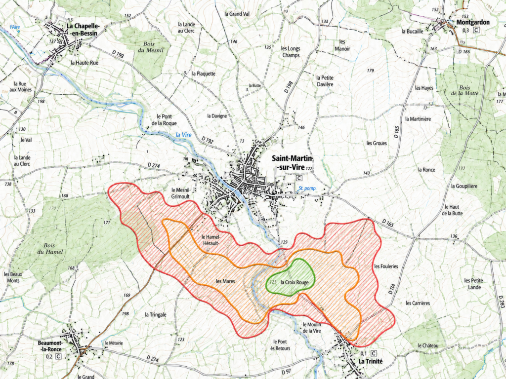
No camera or radar can replace the preparatory work that narrows down the area to be searched. A single grave rarely exceeds two square metres, and searching blindly across hectares of woodland or field makes no sense. Investigators therefore begin with a desk-based study cross-referencing the suspect’s movements, telephone data, witness accounts and aerial imagery, sometimes historical, that may reveal disturbed ground or a vehicle access point [2]. To this is added an assessment of what geologists call diggability, that is, how easily a person could actually open and backfill a grave given soil depth, the local geology, the water table, obstructions or roots, and the tools available [2]. A Red-Amber-Green prioritisation system then ranks sectors by likelihood, so that resources are focused where they are most useful [2].

Système de priorisation par couleurs, rouge, orange et vert, permettant de hiérarchiser les secteurs de recherche selon leur probabilité, afin de concentrer les moyens là où ils sont le plus utiles. Crédit : ForenSeek

This approach was formalised in 2021 as the geoforensic search strategy, which works from the broadest scale down to the smallest area and from non-intrusive to intrusive methods [3]. In practice it behaves like a funnel. Before ever setting foot on the suspect ground, teams calibrate their instruments over reference test graves to learn what the signal looks like under the site’s environmental conditions. Next comes the detection of surface anomalies, supported by cadaver dogs, then non-invasive geophysical methods such as GPR. Only after these stages does targeted probing follow, and finally full excavation of the site [4][5]. This order is not administrative convenience. It avoids destroying a potential scene and reserves heavy resources for genuinely promising areas.

II. Looking from above, remote sensing and drones

Surface relief betrays the grave

The first sign a grave leaves at the surface is topographic. Digging loosens the soil, which then occupies a greater volume than intact ground and forms a slight mound. Over time, settling of the backfill and collapse of the soft tissues during decomposition instead produce a depression. Oblique aerial photography and drone imagery exploit these variations, but they are fleeting and often hidden by vegetation or smoothed away by weather. To recover this micro-relief beneath tree cover, teams turn to LiDAR, a laser scan that records millions of points and reconstructs the bare-earth surface once vegetation is digitally stripped away, revealing subtle depressions or mounds. LiDAR has the advantage of penetrating gaps in the canopy, but it detects a landform rather than the body itself, and its performance drops under very dense cover.

Spectral sensors

Beyond relief, decomposition alters the light signature of soil and plants, which multispectral and hyperspectral sensors measure in bands invisible to the naked eye. The published results call for caution. Most studies can only separate disturbed soil from undisturbed soil, without being able to confirm that a grave holds a body rather than merely turned earth [9]. The frequently cited exception is the study by Kalacska and Bell, who distinguished graves from their controls through weaker vegetation regrowth over the graves, attributing that failure to recover to soil toxicity caused by the leaching of decomposition products [7]. In hyperspectral imaging, work has detected buried remains a few months after burial, including in arid settings, and located single graves [8][9]. A programme in Colombia, a country with more than 120,000 missing people, offers a striking data point, since experimental graves there remained detectable eight years after burial using near-infrared multispectral imaging and NDVI, though not with conventional optical sensors [6]. That index, NDVI or the Normalized Difference Vegetation Index, compares red and near-infrared light to gauge plant vigour, and it illustrates the variability of these methods, having revealed graves in some studies while yielding no usable anomaly in others [9]. A spectral sensor therefore guarantees nothing, and its performance depends closely on elapsed time, climate and soil type.

Thermal imaging

Drone-mounted thermal imaging rests on a simple principle. Disturbed earth differs from surrounding soil in compaction and moisture, hence in how it stores and releases heat, and active decomposition itself gives off a little energy. These temperature contrasts show up on an infrared camera, especially at times of day when the ground warms or cools quickly. Studies have accordingly assessed the detection of clandestine graves by airborne thermal imaging in arid environments [9]. The method’s strength lies in its speed and its ability to cover large areas. Its weakness is that the observation window is narrow and highly condition-dependent, which is why dry, high-contrast settings suit it best.

III. Probing the subsurface, geophysical methods

Ground-penetrating radar

Ground-penetrating radar, or GPR, is the central tool of geophysics applied to criminal investigations. It transmits electromagnetic pulses into the ground and records their echoes, which reveal discontinuities such as a grave cut, a wrapped body or a cavity. Since the mid-1990s it has been used to locate clandestine graves, search for missing persons and investigate mass graves [10]. In 2013, in volcanic tuff caves in Italy, a 500 MHz antenna detected a strong anomaly at 2 metres’ depth corresponding to an air-filled burial cavity, which excavation confirmed to contain human remains [10]. Like other geophysical methods, it has the advantage of being non-destructive, fast and inexpensive, and of avoiding needless disturbance of the remains [11]. Frequency choice governs the result, and the landmark ten-year study is clear on this point. Medium frequencies, from 225 to 450 MHz, offer the best trade-off between resolution, depth of investigation, few false anomalies and speed of acquisition, with any wrapping around the body acting as an excellent reflector [12]. GPR’s main limitation is that it loses much of its effectiveness in clay-rich or highly conductive soils, which then calls for pairing it with other sensors [10].

Electrical resistivity

Electrical resistivity tomography measures how the ground resists the passage of a current, a decomposing body and disturbed soil locally altering that resistance. It often complements GPR and takes over where the latter fails. The same ten-year longitudinal study shows that the signal changes over time, which is crucial for cold cases. A naked burial produces large, low-resistivity anomalies for about four years, after which the body becomes hard to image, whereas a wrapped burial yields small, high-resistivity anomalies for four years, then larger anomalies that remained detectable until the end of the ten-year monitoring period [12]. The same work recommends using resistivity in clay-rich soils, precisely where GPR loses its edge, and running both methods together when it is unknown whether the body was wrapped [12][13]. It also shows that winter and spring surveys offer the best chance of detection [12]. The fact that unsolved cases are typically reviewed roughly every ten years gives such long-term monitoring very concrete relevance for investigators [12].

Other geophysical sensors

Other instruments round out the subsurface toolkit. Bulk-ground electromagnetic conductivity rapidly maps the soil’s overall conductivity and helps track the migration of decomposition fluids, as the same Colombian monitoring showed by combining drone, GPR, resistivity and conductivity over four to eight years [6]. Magnetometry and magnetic susceptibility mainly detect ferromagnetic objects and contrasts in disturbed or burnt soil, so they target items associated with a burial rather than the body itself, though magnetic susceptibility has been proposed as a search tool [14][15]. Metal detectors, finally, remain useful for locating metallic items buried with the victim, such as jewellery, projectiles, tools or binding wire.

IV. When the living reveal the dead, vegetation, fungi and soil chemistry

Disturbed vegetation

A grave affects plant life in two complementary ways. Digging first destroys the vegetation in place, which is then recolonised by ruderal species, the plants that settle first on disturbed ground, forming a patch distinct from the surrounding cover. At the same time, decomposition releases nutrients that may stimulate growth, or conversely compounds in excess that suppress it. An experimental trial using five pig carcasses buried in Italy, with monthly recording of every plant for a year, confirmed that burial alters plant communities through both mechanical disturbance and changes in the nutrient balance [16]. These changes are sometimes subtle but persistent, and they are all the more legible in naturally poor environments, where a sudden nutrient input favours a markedly different flora above the body [16]. One associated chemical marker, the influx of ninhydrin-reactive nitrogen into gravesoil, has indeed been measured during decomposition [19]. This botanical approach, already part of multidisciplinary grave-detection strategies as early as 1992 [17], is today documented by several field studies [18].

Canopy colour, an appealing but fragile lead

Among the botanical clues, the idea that tree canopies or leaf colour might betray a buried body is the most spectacular, and also the one to handle with the greatest care. For now it belongs to exploratory research rather than to a validated investigative method. Researchers have proposed turning plant cover, usually seen as an obstacle, into an asset, by suggesting that plant phenotyping from satellite or drone could detect a massive nitrogen input or stress responses such as chlorosis [20]. According to the authors, the speed at which a plant reacts to this nitrogen influx could change the colour and reflectance of its foliage, yet they immediately note that other large mammals, a deer for instance, also die in the places where people go missing, which opens the door to false positives [20]. At this stage, canopy colour is a promising working hypothesis, to be presented as such and not as an operational technique.

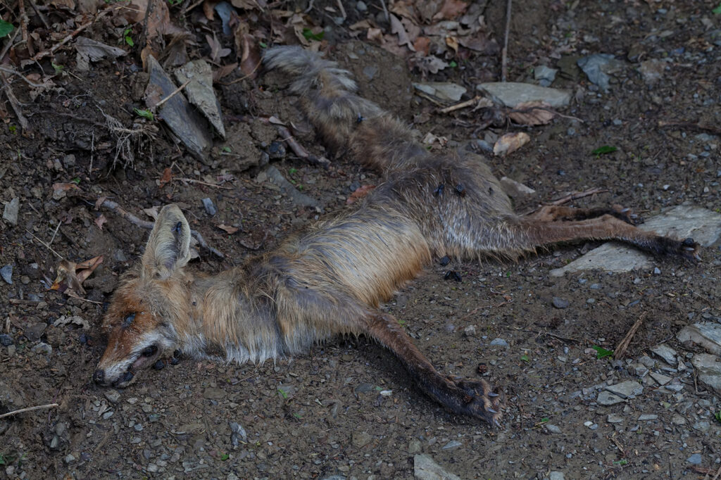
Décomposition d’un renard en zone forestière

Fungi, discreet grave markers

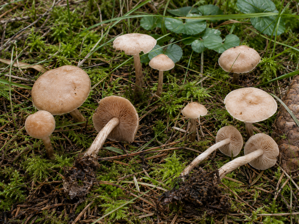
Forensic mycology offers a lesser-known and more surprising clue. Two closely related groups, ammonia fungi and so-called postputrefaction fungi, are associated with the breakdown products of cadavers, and their fruit bodies have been observed in woodlands worldwide, sometimes marking grave sites in characteristic successions [21]. These fungi thrive in soils rich in nitrogen compounds, the ammonia released by decomposition being necessary for their fruiting, which explains their affinity for shallow graves [21]. Some species of the genus Hebeloma have even earned the nickname corpse finder, such as Hebeloma syrjense, whose taxonomic status nonetheless remains debated [21]. Two limitations call for great caution. First, no published criminal case has established that fungi enabled a body to be located, the value of these observations remaining to be demonstrated for casework [23]. Second, the species that colonise the soil around a grave are not the same as those growing directly on the remains, so one must avoid treating any fungus present on a body as a grave marker [21][23]. The lead is real and has been documented since the founding work on ammonia fungi [22], but it remains a supporting tool still under evaluation.

Découverte de champignons Hebeloma syrjense en zone forestière. Crédit : ForenSeek

The cadaver decomposition island and the soil’s chemical signature

Beneath most of these clues lies one and the same phenomenon, the local transformation of soil by decomposition. Scientists speak of the cadaver decomposition island to describe this zone enriched in compounds released by the body, a genuine biochemical hotspot where nitrogen, phosphorus and other elements flood the underlying soil [24]. Recent data from human cadavers quantify this enrichment. A 2025 study, the first of its kind to measure the stable isotopes of carbon and nitrogen in human gravesoils, shows that soil nitrogen content more than doubles between days ten and fifteen, rising from about 0.4 to 1.05 per cent, while the nitrogen isotopic signature, δ15N, becomes enriched by fifteen to twenty per mil over the first month and stays elevated thereafter [25]. Tellingly for investigators, this marker can betray a decomposition island even when the body has been moved [25]. These indicators, nitrogen, phosphorus, acidity, conductivity and isotopes, become usable through sampling once a suspect area has been identified, and they are accompanied by a profound shift in the soil’s microbial communities that tracks the progress of decomposition [24]. This chemical signature is the objective foundation on which botany, mycology and odour detection all rest to varying degrees.

V. Following the odour, dogs and instrumental detection

Human remains detection dogs

In the field, the dog remains one of the most effective ways to locate remains. These animals are trained to detect the volatile organic compounds given off by decomposition, the odorous molecules that diffuse through soil and air. The measured performance is notable. On gravesoils, dogs gave correct responses in close to 93 per cent of trials, and they detected the presence of remains in soil up to 915 days after death, with the oldest sample identified in 100 per cent of cases [26]. An earlier field programme had established a recovery rate of about 81 per cent [27], and chemical analysis has identified several signature compounds above human burials [28]. Soil texture nonetheless influences the outcome, since it governs the escape of decomposition gases, a sandy soil allowing a faster response than a clay one [29]. These strengths come with limitations that must be acknowledged. Trained dogs are costly to train and maintain, work only for short periods, can give false alerts and cannot indicate precisely what they are detecting [30].

The electronic nose

To make this sense of smell objective and to ease its constraints, research is developing electronic noses able to analyse volatile compounds. A portable device, the NOS.E, detected and differentiated a range of decomposition-related molecules with an average sensitivity of around eight parts per million, and told a human donor from controls as early as the third day after death, while offering portability, speed and lower costs than dogs and benchtop instruments [31]. The most recent approaches couple sensor arrays with machine learning, a thirty-two metal-oxide-sensor electronic nose having classified post-mortem versus ante-mortem samples with 98.1 per cent accuracy and distinguished human from animal tissue with 97.2 per cent accuracy [32]. The practical limitation lies in odour dispersal, since a stationary device may miss a diluted plume, and in the sampled matrix, since soil and air do not yield exactly the same compounds, which argues for sampling both [31][33]. The electronic nose does not yet replace the dog, but it makes for a promising and reproducible complement.

VI. From anomaly to evidence, confirmation and recovery

None of these non-intrusive methods proves on its own that a body is present, since all of them flag an anomaly that must be confirmed. After GPR come targeted probing, carried out by coring or with a metal probe whose escaping gases are smelled, then sampling of soil, vegetation or water, before progressive stripping of the ground and full excavation [4]. This last stage falls to forensic archaeology and anthropology, the only disciplines able to expose the remains by a rigorous method that preserves their position, the associated traces and the evidential value of the whole [34]. It is this controlled passage from a detected anomaly to a documented exhumation that turns a hypothesis about location into evidence usable before a court.

Comparative summary of methods

The table below summarises the main methods, what they aim to detect, their favourable conditions, their chief limitation and how intrusive they are.

MethodWhat it targetsFavourable conditionsMain limitationIntrusiveness
Aerial photography, micro-reliefGround surface disturbanceOpen ground, recent graveFleeting traces, hidden by vegetationNone
LiDARMicro-depressions under coverWooded areas, bare-earth modellingDetects landform, not the bodyNone
Multispectral and hyperspectral imagingPlant stress, disturbed soilAfter regrowth, a few months onVariable results, sensitive to bare soilNone
Drone thermal imagingThermal contrast of disturbed soilArid settings, diurnal transitionsNarrow window, highly condition-dependentNone
Ground-penetrating radarGrave cut, body, cavitySandy or dry soils, wrapped bodyFails in clay-rich or conductive soilsNone
Electrical resistivitySoil resistivity anomalyHomogeneous soil, moist conditionsSignal evolves and fades over timeNone
Electromagnetic conductivityDecomposition fluids, disturbed soilRapid mapping of large areasLower resolution than resistivityNone
Magnetometry, susceptibilityFerrous objects, burnt soilAssociated metallic itemsTargets the object more than the bodyNone
Vegetation, botanyShift in plant communityPoor soils, monitoring over timeSubtle, non-specific clueLow
Canopy colour (phytoforensics)Induced reflectance and chlorosisConcept, yet to be validatedUnvalidated, false positives (wildlife)None
Marker fungiFruit bodies tied to decompositionWoodlands, seasonal fruitingNo judicial validation, transient presenceLow
Soil chemistry, decomposition islandNitrogen and isotope signatureAlready pre-selected areaRequires targeted samplingLow
Detection dogsVolatile decomposition compoundsLoose soil, short sessionsCost, fatigue, false alerts, non-explainable resultNone
Electronic noseObjectified volatile compoundsSoil and air samplingPlume dispersal, immature technologyLow
Probing, forensic archaeologyConfirmation and recoveryConfirmed restricted areaDestructive, slowHigh

Conclusion

Searching for a buried body is therefore not a matter of some miracle sensor, but of a coherent chain in which intelligence delimits the ground, remote sensing and geophysics flag anomalies, botany, mycology and soil chemistry provide converging clues, and excavation confirms. Success always depends on soil type, climate, depth, elapsed time and the way the body was buried, variables that shift the real question from which method to use towards which method to use, when, and in which soil. In a case such as that of Delphine Jubillar, a confession and an indication of place do not close the search, they reopen it, and it is precisely this scientific arsenal that will have to turn a statement into a location, and a location into remains at last returned to their family.

Références :

[1] Affaire Jubillar, de la disparition de Delphine aux aveux de Cédric. CNews, 6 juillet 2026. Aveux confirmés par La Dépêche du Midi et l’Agence France-Presse.

[2] Harrison M, Donnelly LJ. Locating concealed homicide victims, developing the role of geoforensics. In Criminal and Environmental Soil Forensics. Dordrecht, Springer, 2009, p. 197-219.

[3] Geoforensic methods for detecting clandestine graves and buried forensic objects in criminal investigations, a review. Journal of Forensic Science and Medicine, 2024, vol. 10, n° 3.

[4] Pringle JK. Geoforensic search on land. Geology Today, 2024, vol. 40, n° 4.

[5] Pringle JK, Ruell A, Jervis JR, Donnelly LJ, McKinley J, Hansen JD, et al. The use of geoscience methods for terrestrial forensic searches. Earth-Science Reviews, 2012, vol. 114, p. 108-123.

[6] Molina CM, Wisniewski KD, Salamanca A, Saumett M, Rojas C, Gómez H, Baena A, Pringle JK. Monitoring of simulated clandestine graves of victims using UAVs, GPR, electrical tomography and conductivity over 4-8 years post-burial to aid forensic search investigators in Colombia, South America. Forensic Science International, 2024, vol. 355, article 111919.

[7] Kalacska M, Bell LS. Remote sensing as a tool for the detection of clandestine mass graves. Canadian Society of Forensic Science Journal, 2006, vol. 39, n° 1, p. 1-13.

[8] Leblanc G, Kalacska M, Soffer R. Detection of single graves by airborne hyperspectral imaging. Forensic Science International, 2014, vol. 245, p. 17-23.

[9] A review of predictive modelling and drone remote sensing technologies as a tool for detecting clandestine burials. Forensic Science International, 2025.

[10] Ground penetrating radar in forensic science, applications, methodologies, challenges, and future directions, a comprehensive review. Perspectives in Legal and Forensic Sciences, 2026.

[11] Pringle JK, Jervis JR, Hansen JD, et al. Geophysical monitoring of simulated clandestine graves using electrical and ground-penetrating radar methods, 0-3 years after burial. Journal of Forensic Sciences, 2012, vol. 57, n° 6, p. 1467-1486.

[12] Pringle JK, Stimpson IG, Wisniewski KD, et al. Geophysical monitoring of simulated homicide burials for forensic investigations. Scientific Reports, 2020, vol. 10, article 7544.

[13] Pringle JK, Jervis JR, Roberts D, et al. Long-term geophysical monitoring of simulated clandestine graves using electrical and ground penetrating radar methods, 4-6 years after burial. Journal of Forensic Sciences, 2016, vol. 61, n° 2, p. 309-321.

[14] Pringle JK, Giubertoni M, Cassidy NJ, et al. The use of magnetic susceptibility as a forensic search tool. Forensic Science International, 2015, vol. 246, p. 31-42.

[15] Juerges A, Pringle JK, Jervis JR, Masters P. Comparisons of magnetic and electrical resistivity surveys over simulated clandestine graves in contrasting burial environments. Near Surface Geophysics, 2010, vol. 8, p. 529-539.

[16] Caccianiga M, Bottacin S, Cattaneo C. Vegetation dynamics as a tool for detecting clandestine graves. Journal of Forensic Sciences, 2012, vol. 57, n° 4, p. 983-988.

[17] France DL, Griffin TJ, Swanburg JG, et al. A multidisciplinary approach to the detection of clandestine graves. Journal of Forensic Sciences, 1992, vol. 37, n° 6, p. 1445-1458.

[18] Watson CJ, Forbes SL. An investigation of the vegetation associated with grave sites in southern Ontario. Canadian Society of Forensic Science Journal, 2008, vol. 41, n° 4, p. 199-207.

[19] Van Belle LE, Carter DO, Forbes SL. Measurement of ninhydrin reactive nitrogen influx into gravesoil during aboveground and belowground carcass (Sus domesticus) decomposition. Forensic Science International, 2009, vol. 193, p. 37-41.

[20] Plants to remotely detect human decomposition ? Trends in Plant Science, 2020, vol. 25, n° 10.

[21] Tibbett M, Carter DO. Mushrooms and taphonomy, the fungi that mark woodland graves. Mycologist, 2003, vol. 17, n° 1, p. 20-24.

[22] Sagara N. Ammonia fungi, a chemoecological grouping of terrestrial fungi. Contributions from the Biological Laboratory, Kyoto University, 1975, vol. 24, p. 205-276.

[23] Hawksworth DL, Wiltshire PEJ. Forensic mycology, the use of fungi in criminal investigations. Forensic Science International, 2011, vol. 206, p. 1-11.

[24] Carter DO, Yellowlees D, Tibbett M. Cadaver decomposition in terrestrial ecosystems. Naturwissenschaften, 2007, vol. 94, n° 1, p. 12-24.

[25] Miles KL, Gibbon V, Hayden B. Human cadaver decomposition islands and forensic taphonomy, gravesoil δ13C and δ15N enrichment patterns in short (30 d) and extended (900 d) postmortem intervals. Forensic Sciences Research, 2025, DOI 10.1093/fsr/owaf027.

[26] Alexander MB, Hodges TK, Bytheway J, Aitkenhead-Peterson JA. Application of soil in forensic science, residual odor and human remains detection dogs. Forensic Science International, 2015, vol. 249, p. 304-313.

[27] Komar D. The use of cadaver dogs in locating scattered, scavenged human remains. Journal of Forensic Sciences, 1999, vol. 44, n° 2, p. 405-408.

[28] Vass AA, Smith RR, Thompson CV, et al. Odor analysis of decomposing buried human remains. Journal of Forensic Sciences, 2008, vol. 53, n° 2, p. 384-391.

[29] Alexander MB, Hodges TK, Wescott DJ, Aitkenhead-Peterson JA. The effects of soil texture on the ability of human remains detection dogs to detect buried human remains. Journal of Forensic Sciences, 2016, vol. 61, n° 3, p. 649-655.

[30] Cadaver-detection dogs, a review of their capabilities and the volatile organic compound profile of their associated training aids. WIREs Forensic Science, 2021.

[31] Sunnucks EJ, Thurn B, Brown AO, Zhang W, Liu T, Forbes SL, Su S, Ueland M. Performance of a novel electronic nose (NOS.E) for the detection of volatile organic compounds relating to starvation or human decomposition post-mass disaster. Sensors, 2024, vol. 24, n° 18, article 5918.

[32] Shtepliuk I, et al. Adaptive machine learning for electronic nose-based forensic volatile organic compound classification. Advanced Science, 2025, DOI 10.1002/advs.202504657.

[33] Decomposition odour profiling in the air and soil surrounding vertebrate carrion, 2014 (référence à confirmer sur la source primaire).

[34] Blau S, Sterenberg J. The use of forensic archaeology and anthropology in the search and recovery of buried evidence. In Encyclopedia of Forensic and Legal Medicine, 2e éd., Elsevier, 2015, p. 236-245.

Iris identification post-mortem avec utilisation en police scientifique - Forenseek

Post-Mortem Iris Identification

For a long time, the iris was thought to become unusable almost as soon as a person died. Several recent studies show the opposite, and the most comprehensive of them has just been published in the United States.

The iris is the coloured ring that surrounds the pupil. Its relief, made up of crypts, furrows and striations, forms a texture that belongs to one individual alone and becomes fixed in early childhood, never changing afterwards. A high-resolution photograph is enough to capture it, after which an algorithm encodes it into a template unique to that person and compares it against every record already held in a database. In France, iris biometrics remains very little used, yet for years it has supported the identification of living individuals elsewhere in the world. In the United States, the FBI has turned it into a service in its own right, the NGI Iris Service, fed by a national network in which sheriffs enrol the iris of suspects and convicted offenders at the time of booking. One question nonetheless remained open, one that almost no one had seriously examined. What becomes of this identifier once the person has died?

A certainty that science eventually overturned

When death occurs, the pupil becomes fixed, most often wide open, and the cornea is gradually veiled by a whitish film, the milky sheen sometimes seen in the gaze of the dead. As early as 2001, John Daugman, the British engineer behind automated iris recognition, told the BBC that this twofold change made the task considerably harder. The notion took firm hold, to the point of feeding the claim, repeated as though self-evident, that the iris became unusable within minutes.

The past 15 years have patiently dismantled that certainty, one study after another [3]. As early as 2016, it was established that an iris could still be recognised several days after death [3]. How the body is preserved soon proved to be the decisive factor. Left outdoors, the iris degrades quickly, whereas fingerprints and the face withstand decomposition far better, as Bolme and colleagues observed [3]. At the other extreme, Sauerwein and colleagues recovered irises that were perfectly analysable 34 days after death, on bodies left outdoors but exposed to the cold of winter [3]. The first studies genuinely tracked over time, carried out in Warsaw by Mateusz Trokielewicz and colleagues, reached a conclusion that few specialists had anticipated. Under favourable conditions, the iris can be analysed without notable difficulty 5 to 7 hours after death, and identification sometimes remains possible up to 3 weeks afterwards [4].

The most comprehensive study to date

The most accomplished demonstration is very recent. It comes from a large team bringing together the University of Notre Dame and Michigan State University, led by Rasel Ahmed Bhuiyan, and it has been accepted by a leading biometrics journal [1]. The researchers first assembled an image collection without precedent, more than 10,000 iris photographs gathered from 259 deceased subjects, in visible light as well as near-infrared, the wavelength that best reveals the texture of the iris [1]. For some of them, the interval between death and image capture reached 1,674 hours, close to 70 days [1].

The corpus includes a case never before published, that of a person whose eye was photographed both before and after death, which made it possible to compare the iris of the living individual directly with that of the deceased [1]. It also contains atypical configurations, rarely documented until then, such as an eye dislodged from its orbit by an injury [1]. Adding the few collections already available, the team brought together images of 338 deceased subjects, the largest sample ever assembled on the subject, then ran them through 5 recognition systems, from the oldest to the most recent, some based on artificial intelligence and others already on the market [1]. The results confirm and refine what the literature had hinted at. When the body has been kept in good conditions, current systems still recognise the iris several hours, sometimes several days, after death [1]. Here again, everything hinges on preservation, and the gap can be considerable. A body left in a field at the height of summer will see its iris degrade within a few days, until any comparison becomes impossible. The same body placed in a refrigerated mortuary drawer at a medico-legal institute, at around 6 degrees Celsius, keeps a usable iris for several weeks [1].

For an investigator, a magistrate or a forensic pathologist, the consequence is clear. No fixed deadline marks the moment when the iris would cease to be identifiable. There are only more or less favourable conditions, which will have to be reconstructed and documented in each case.

The study does not stop at measuring performance. It also delivers an open-source software tool, named PMExpert, designed to support the examiner and already in use since 2021 within a United States medico-legal institute [1]. Rather than returning a definitive ‘yes’ or ‘no’, it highlights the regions of the iris on which it relies to match two images, ante-mortem and post-mortem, leaving the expert free to verify and weigh the correspondence [1]. The software and the accompanying data are made available to forensic services free of charge, through the public United States criminal justice data archive [1].

Estimating how long a person has been dead

The same images fed a second piece of research, one that must be carefully distinguished from the first [2]. This one no longer seeks to identify an individual from the iris, but to estimate how long that individual has been dead. Conducted by Rasel Ahmed Bhuiyan and Adam Czajka and presented at the WACV 2025 conference, it draws on European collections and on the new dataset, 348 subjects in all [2]. The principle is nothing new. For centuries, the forensic pathologist has estimated the post-mortem interval from the positive signs of death that evolve at a relatively steady pace, such as algor mortis, the onset of livor mortis, or the appearance and then the disappearance of rigor mortis. What is new lies in the sign chosen and in the way it is read.

Here, what the machine observes is the white veil that gradually spreads across the eye after death. The more advanced it is, the longer ago death occurred. The researchers trained a deep learning model to perform this reading from the iris photograph alone [2]. Accuracy depends, once again, on environmental factors. In the scenario closest to real conditions, when the model must rule on bodies and settings it has never encountered, it is off by an average of about 3 days [2]. It can therefore point to a window, but never to a precise hour. Feasibility is established, yet we remain far from a fully reliable tool for dating death [2].

For the courts, a lead to handle with caution

In practical terms, what use is this in an investigation? Faced with an unidentified body, the eye is photographed, the software proposes a list of possible matches from a database, and an expert then confirms or rules out each of them [1]. The iris will replace neither DNA, nor fingerprints, nor forensic odontology, the three primary identifiers used to formally identify a body. It is an addition to them, above all when those methods take time or prove unusable, for want of ante-mortem data, on bodies that are too degraded or fragmented.

Its main advantage is speed. Comparing the iris of an eye against a biometric database takes only a few seconds, an argument that can weigh heavily when many victims must be identified under time pressure, after a mass-casualty event for example. This ability to reliably identify someone after death nonetheless raises a security concern of the first order. If the iris of a deceased person remains identifiable, one can imagine that a deliberately removed eye, or even a stored image, might be used to unlock a phone or to clear a biometric checkpoint based on this method. The authors anticipated this scenario and adapted software designed to flag fake eyes, a form of presentation attack detection, showing that it very quickly learns to recognise a deceased person’s iris and reject it [1].

Conclusion

The real reach of this advance remains to be measured. The window of opportunity exists, but it closes quickly, as the corneal veil thickens and the tissues decompose. The images needed to train and test the software remain scarce and hard to gather, something the researchers themselves acknowledge as a major obstacle [1]. And no framework yet defines what would make such an identification admissible in court. As long as that framework is missing, the iris of a deceased person is to be weighed like any other expert finding, with its conditions to document and its share of debate.

One obvious point deserves to be stressed above all. Identifying someone from the iris presupposes that this iris is already on file, recorded while the person was alive, when applying for a biometric passport in countries that provide for it, or when gaining access to a secure site through iris recognition. Without that reference, even the most powerful analysis leads nowhere. In France, where the iris feeds no identification file, the technique is therefore, for now, a matter of horizon-scanning more than routine practice. It nonetheless deserves close attention, for it illustrates a deeper trend, that of a forensic science learning to make the body speak when DNA and fingerprints fall silent.

References

  • [1] Bhuiyan R. A., Farmanifard P., Sharma R., Kuehlkamp A., Boyd A., Flynn P. J., Bowyer K. W., Ross A., Chute D., Czajka A. (2026). Beyond Mortality, Advancements in Post-Mortem Iris Recognition through Data Collection and Computer-Aided Forensic Examination. IEEE Transactions on Biometrics, Behavior, and Identity Science, early access. arXiv:2603.26976. https://ieeexplore.ieee.org/document/11063436
  • [2] Bhuiyan R. A., Czajka A. (2025). Forensic Iris Image-Based Post-Mortem Interval Estimation. IEEE/CVF Winter Conference on Applications of Computer Vision, WACV 2025. arXiv:2404.10172.
  • [3] Boyd A., Yadav S., Swearingen T., Kuehlkamp A., Trokielewicz M., Benjamin E., Maciejewicz P., Chute D., Ross A., Flynn P., Bowyer K., Czajka A. (2020). Post-Mortem Iris Recognition, A Survey and Assessment of the State of the Art. IEEE Access, vol. 8, p. 136570-136593.
  • [4] Trokielewicz M., Maciejewicz P., Czajka A. (2024). Post-mortem Iris Biometrics, Field, Applications and Methods. Forensic Science International.

Analyse ADN laboratoire police scientifique transfert

When an absent investigator’s DNA turns up on a sealed exhibit

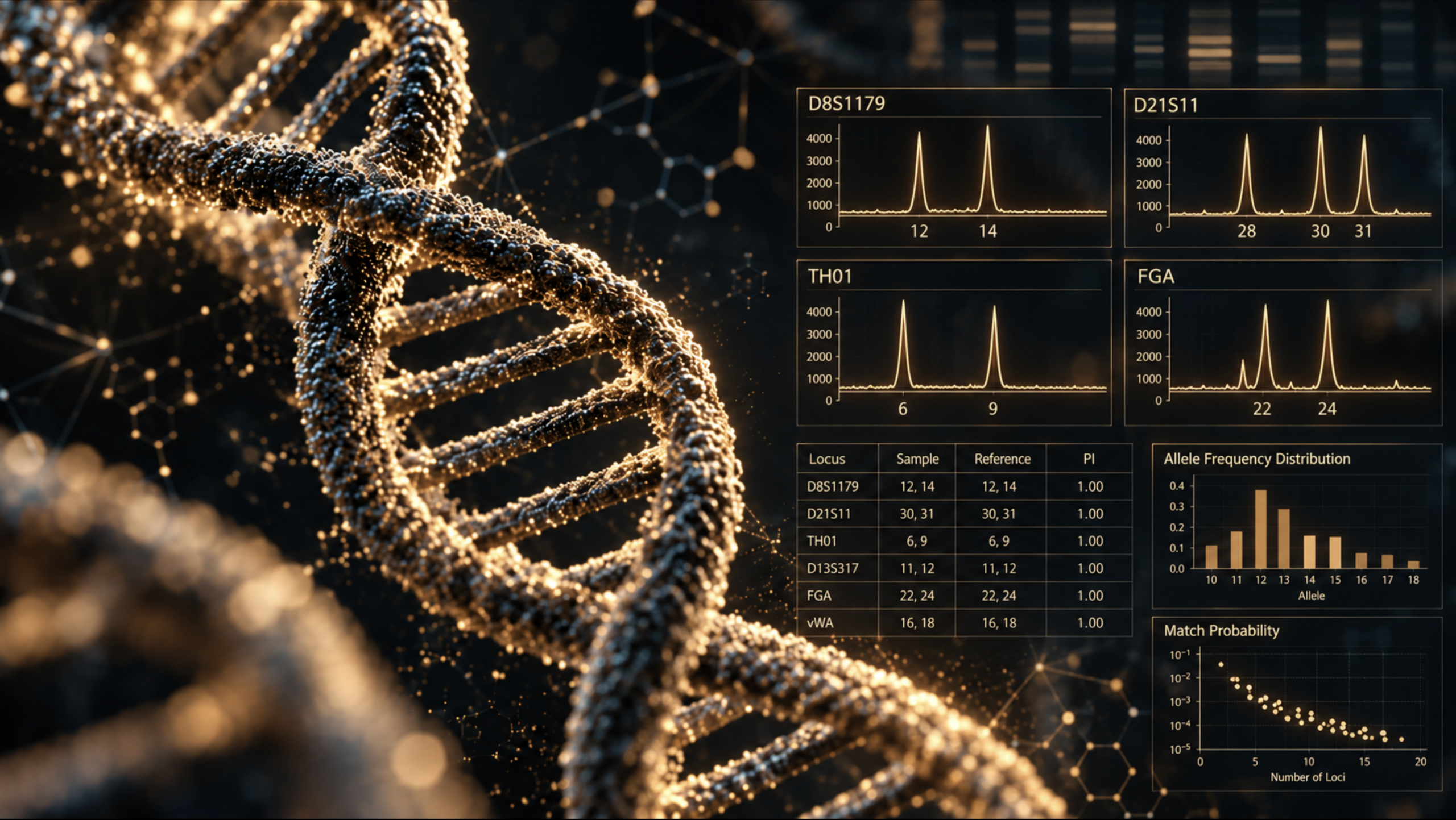
A case that had gone unsolved for eighteen years is reopened. From a plastic bag kept as a sealed exhibit, analysts obtain a full DNA profile. Run against the staff elimination database, it returns a name, that of an investigator. The trouble is that this investigator was eighty kilometres from the laboratory and had never set foot at the scene or in the exhibit storage room. The profile was nonetheless his. DNA had identified a person with near-certainty on an item he had never handled. Reported in early 2026 by Gao et al. in Forensic Science International: Genetics, this case is a reminder of a distinction that is too often overlooked: knowing whose DNA a sample carries and knowing how it got there are two different questions.

Identifying the source is not explaining the activity

The forensic laboratory’s result answered one question perfectly, the origin of the genetic material, but it offered no reliable information about the circumstances of deposition. This distinction has long shaped the reasoning of forensic geneticists. In 1998, Cook, Evett et al. [1], writing in Science & Justice, proposed a hierarchy of propositions setting out three levels of interpretation, running from the source to the offence itself. The sub-source level, specific to DNA, was added later, notably by Evett et al. in 2002 [2]. Establishing whose biological profile a sample matches sits at the very bottom of this ladder. Explaining how that profile came to be there, on a substrate or at a crime scene, belongs to the activity level, which is higher and far harder to establish. For a judge, the distinction is decisive. A laboratory can state with great confidence that the DNA profile obtained from a biological sample matches an individual’s profile, yet cannot say, with the same assurance, what that presence means.

The four levels of interpretation: a textbook case

The classic Cook and Evett example, a sexual assault case, where each level adds a further layer of inference on top of the one below:

  • Sub-source level:
    “The DNA in the sample comes from the suspect” (rather than from an unknown person). This is what the raw DNA comparison measures directly. It speaks only to DNA, nothing more.
  • Source level:
    “The semen comes from the suspect.” This adds identification of the body fluid, provided it has been established that the stain really is semen.
  • Activity level:
    “The suspect had sexual intercourse with the complainant.” This adds the mechanism of deposition where transfer, persistence and contamination come into play.
  • Offence level:
    “The suspect raped the complainant.” This adds the absence of consent which the expert cannot decide, since that is for the court.

At each step up, science can say a little less, and the weight of the evidence diminishes.

How more sensitive methods changed the picture

The problem is not new, but it has changed in scale as DNA analysis has advanced. Today’s kits can recover usable DNA profiles from just a few cells, where once a far larger amount of DNA was needed. In 1997, van Oorschot and Jones [9] showed that mere contact is enough to deposit DNA. Since then, the literature has documented many routes of transfer, both direct and indirect, surveyed in van Oorschot et al.’s 2019 review [10]. The effect cuts both ways. The very sensitivity that makes it possible to reopen old cases also raises the chance of detecting a DNA profile unrelated to the events in question. Studies remain cautious about frequencies, which vary widely with conditions, so much so that some authors regard secondary transfer as an uncommon event. Generalisation must therefore be handled with care, in both directions.

The risk of DNA transfer in evidence handling

The journey of a sealed exhibit, from the crime scene to the laboratory bench, passes through a series of handling steps during which DNA can move. The packaging of the item itself plays a part. Stella et al. [7] showed in 2026 that this transfer can run two ways: DNA deposited on an item may migrate to the inner surface of the packaging, so that the sought-after profile is missing from the analysis; or it may transfer to another area of the item, where it has no business being and clouds interpretation. Goray et al. [6] noted in 2019 that the outer surface of gloves could carry, alongside the target profile, that of the examiners themselves, particularly when opening and resealing the packaging. It is precisely this risk that the examination protocols used by forensic services are designed to head off, requiring decontaminated gloves changed for each new item. Examination tools pose a comparable problem, described as early as 2015 by Szkuta et al. [8]. That an investigator might contaminate an item or surface after the fact is therefore no far-fetched hypothesis; Fonneløp’s team [3,4] has studied the phenomenon right down to evidence bags themselves.

To explain the presence of this profile on an exhibit the investigator had never come near, Gao et al. [5] rule out item-to-item transfer in favour of a likely airborne route: the DNA is thought to have settled on a colleague’s clothing before detaching as that colleague approached the sample pre-treatment area. Without the staff elimination database, this hypothesis would never have come to light.

What this distinction changes for the evaluation of evidence

For legal professionals, the lesson of this case is not that DNA is unreliable, but rather that a solid genetic identification at the source level can coexist with a fragile explanation at the activity level. The whole risk lies in sliding from one to the other, when the strength of the former colours the assessment of the latter. To guard against this slide, some forensic geneticists argue that the expert should reason explicitly at the activity level and set out the scenarios consistent with the observations. This is notably the position of the School of Criminal Justice at the University of Lausanne and of the European guideline on evaluative reporting [11].

Conclusion :

The case of the investigator who was never at the scene illustrates the stakes better than any theoretical reminder could. His DNA profile, entirely genuine, sat on a sealed exhibit he had never touched, in a case whose scene he had never visited. Without a safeguard that brought another explanation to light, this finding could have been read as a genuine investigative lead backed by a “relevant” DNA profile. A genetic result is worth not only what it identifies, but what it reasonably allows one to infer, and that is never settled on a single trace alone. A DNA profile must be weighed within the wider body of evidence, never in isolation.

References :

  • [1] Cook R., Evett I.W., Jackson G., Jones P.J., Lambert J.A. (1998). A hierarchy of propositions: deciding which level to address in casework. Science & Justice, 38(4), 231-239.
  • [2] Evett I.W., Gill P.D., Jackson G., Whitaker J., Champod C. (2002). Interpreting small quantities of DNA: the hierarchy of propositions and the use of Bayesian networks. Journal of Forensic Sciences, 47(3), 520-530.
  • [3] Fonneløp A.E., Egeland T., Gill P. (2015). Secondary and subsequent DNA transfer during criminal investigation. Forensic Science International: Genetics, 17, 155-162.
  • [4] Fonneløp A.E., Johannessen H., Egeland T., Gill P. (2016). Contamination during criminal investigation: Detecting police contamination and secondary DNA transfer from evidence bags. Forensic Science International: Genetics, 23, 121-129.
  • [5] Gao L. et al. (2026). A rare and atypical case of long-distance indirect DNA transfer: Contamination from an investigator never present at the scene. Forensic Science International: Genetics.
  • [6] Goray M., Pirie E., van Oorschot R.A.H. (2019). DNA transfer: DNA acquired by gloves during casework examinations. Forensic Science International: Genetics, 38, 167-174.
  • [7] Stella C.J., Goray M., Meakin G.E., van Oorschot R.A.H. (2026). DNA transfer in packaging: Investigation of mitigation strategies. Journal of Forensic Sciences, 71(1), 197-210.
  • [8] Szkuta B., Harvey M.L., Ballantyne K.N., van Oorschot R.A.H. (2015). DNA transfer by examination tools, a risk for forensic casework? Forensic Science International: Genetics, 16, 246-254.
  • [9] van Oorschot R.A.H., Jones M.K. (1997). DNA fingerprints from fingerprints. Nature, 387, 767.
  • [10] van Oorschot R.A.H., Szkuta B., Meakin G.E., Kokshoorn B., Goray M. (2019). DNA transfer in forensic science: A review. Forensic Science International: Genetics, 38, 140-166.
  • [11] ENFSI, Guideline for Evaluative Reporting in Forensic Science, 2015

arnaques sentimentales brad pitt enquête forenseek police scientifique

How do romance scams work?

Understanding the effectiveness of romance scams

Romance scams rely on robust psychological mechanisms that have been widely studied in criminology and social psychology. Their effectiveness does not stem from individual vulnerability, but from a gradual construction process. The interaction often begins in a mundane way before evolving into a personalized and coherent relationship. The offender adapts their discourse, adjusts response timing, introduces credible biographical elements, and progressively establishes a climate of trust.

This process is based on a gradual commitment of the victim to the relationship, a narrative coherence that reduces the perception of inconsistencies, the mobilization of emotions such as attachment, concern, and urgency, and the emergence of cognitive dissonance when initial doubts arise.

The turning point: cognitive dissonance

Cognitive dissonance represents a critical stage, and likely one of the most decisive mechanisms in the persistence of the fraud. It reflects an internal tension between two incompatible realities: on one side, an emotionally invested relationship, and on the other, the possibility of deception. Acknowledging the scam involves far more than a rational reassessment. It means admitting an error, often financially costly, but above all emotionally significant.

At this stage, several barriers emerge. Feelings of guilt, the realization of having been manipulated, fear of judgment or misunderstanding from relatives, and the perception of having reached a point of no return all contribute to maintaining the relationship. This is compounded by a form of abrupt disillusionment that can be psychologically difficult to process.

In this context, continuing the interaction may paradoxically serve as a way to preserve internal consistency and avoid a significant emotional shock. It is precisely at this moment that disengagement becomes the most difficult, even though warning signs are the most evident. As the relationship progresses, decisions are no longer evaluated purely on a rational basis. They are embedded in an already invested relational framework, which explains why informed and socially integrated individuals can also become victims.

From interaction to trace

From a forensic perspective, these cases generate specific types of traces. Unlike traditional material evidence, the available elements consist primarily of digital interactions.

Written messages, audio recordings, images, and videos form a dataset that documents not only events but also an interaction. These elements allow for the analysis of the offender’s discourse structure, the rhythm of solicitations, the turning points leading to financial requests, and the strategies used to adapt to the victim’s reactions. The introduction of manipulated content, particularly through deepfake techniques, further complicates this analysis. Such materials enhance perceived credibility without constituting direct proof of identity. In practice, meaning does not emerge from isolated elements, but from how they connect and unfold over time.

Objectives and constraints of the investigation

Investigations into romance scams pursue several simultaneous objectives.

  • The first concerns financial flows, with the aim of tracing transactions, identifying intermediary accounts, and, where possible, initiating asset recovery procedures.
  • The second focuses on identifying the perpetrators. In many cases, these scams involve organized networks, with a division of roles between profile creators, conversation operators, and financial intermediaries.
  • The third objective is to understand the modus operandi in order to support prevention efforts and improve detection capabilities.

These investigations are made particularly complex by several factors, including geographical fragmentation, the use of multiple identities, and reliance on dispersed technical infrastructures.

The central challenge of interpretation

One of the main challenges lies in interpreting the exchanges. Like any trace, digital communications only make sense within their context. A message cannot be understood independently of the relationship in which it occurs. It reflects not only the offender’s actions but also how the victim responds under the influence of manipulation. This requires a particularly rigorous analytical approach. The analysis must account for the chronology of interactions, the emotional context, identifiable manipulation strategies, and corroborating external elements. Without this contextualization, the risk of misinterpretation remains significant.

Towards a forensic science of interaction ?

These cases illustrate a broader evolution in the types of traces used in forensic science. Alongside traditional material and digital evidence, interactional traces are emerging, generated through technology-mediated relationships. Deepfakes, fake profiles, and scripted conversations do not represent a fundamental rupture. They extend existing manipulation strategies while increasing their credibility and scalability. The challenge for forensic science lies in integrating these new forms of traces into robust methodological frameworks capable of combining technical data with behavioral analysis.

Judicial challenges and perspectives

For legal practitioners, these cases raise specific issues. The legal qualification of the facts, the assessment of victim consent, and the evidentiary value of exchanges require a nuanced approach.

A first obstacle lies in underreporting. Victims still rarely file complaints, often due to shame, guilt, or fear of judgment. Data from the Federal Bureau of Investigation indicate that reported losses represent only a fraction of the actual phenomenon. Even when cases are reported, proceedings rarely lead to successful prosecutions. The transnational nature of these crimes, the use of fictitious identities, and the complexity of financial flows significantly limit both prosecutions and convictions.

Although data are abundant, their interpretation requires particular rigor. The objective is not to accumulate evidence, but to produce a coherent and scientifically grounded analysis. These developments call for stronger collaboration between investigators, analysts, and behavioral science experts in order to better address offences where evidence lies as much in interaction as in technical data.

Sources

Federal Trade Commission (2024). Consumer Sentinel Network Data Book 2023 – Romance scam losses. Available online:https://www.ftc.gov/reports/consumer-sentinel-network-data-book-2023

Whitty, M. T., & Buchanan, T. (2022). The online romance scam: Causes and consequences. Crime Science. Available online: https://crimesciencejournal.biomedcentral.com/articles/10.1186/s40163-022-00166-2

France Bleu (2025). Retour sur l’arnaque au faux Brad Pitt qui a coûté 830 000 euros à une internaute française. Available online: https://www.francebleu.fr/infos/faits-divers-justice/retour-sur-l-arnaque-au-faux-brad-pitt-qui-a-coute-830-000-euros-a-une-internaute-francaise-9633205

Le Monde (2025). Qui se cache derrière les arnaques sentimentales qui se multiplient en France ?
Available online: https://www.lemonde.fr

Federal Bureau of Investigation (2024). Internet Crime Report – Romance scams.
Available online: https://www.ic3.gov/Media/PDF/AnnualReport/2023_IC3Report.pdf

Can human activity be reconstructed from a smartphone ?

A recent study conducted by researchers at the Netherlands Forensic Institute and the University of Amsterdam examines whether data generated by smartphone sensors can be meaningfully exploited in a judicial context. The central question is whether motion signals recorded by these devices can be translated into a probabilistic evaluation capable of assessing whether a specific human activity occurred at a particular point in time. As everyday objects increasingly capture fine-grained behavioral traces, the methodological integration of such data into judicial reasoning has become a critical issue.

Understanding Human Activity Recognition

Human Activity Recognition, commonly referred to as HAR, is a research field that has developed over the past decade, initially within connected health, sports tracking, and smart device applications. Its principle is to use embedded smartphone sensors to identify categories of movement based on numerical signals. Most smartphones are equipped with an accelerometer and a gyroscope that continuously record variations in movement and orientation. From these time series, statistical models trained on known activities learn to recognize characteristic patterns. Walking, running, and periods of inactivity generate distinct signatures that the algorithm can compare with data extracted from a phone under examination.

In everyday use, this classification supports functions such as physical activity tracking or fall detection. In a judicial setting, however, the objective is narrower and more demanding. The issue is not simply to label a movement, but to determine what these recordings can reasonably support regarding a specific activity, and what they cannot establish.

From classification to probabilistic evaluation

In forensic science, the task is not to assert that an event occurred, but to assess the strength with which observed data support one hypothesis over an alternative. Outputs from activity recognition models can be incorporated into this framework by expressing their evidential value in the form of a likelihood ratio. In practical terms, this involves evaluating whether the recorded sequence is more probable under one clearly defined scenario than under another. The algorithm does not decide that a person was running or walking. It provides a comparative measure of how strongly the data support one of the competing hypotheses.

Methodological illustration in a judicial context

Consider a smartphone seized in the course of an assault investigation. At a time considered critical to the events, the motion sensors recorded a sequence of data.

Two hypotheses are formulated. Under the first, the individual was walking normally. Under the second, the individual was running. The analysis indicates that the observed signals are ten times more probable under the running hypothesis than under the walking hypothesis. The likelihood ratio is therefore ten in favor of the proposition that the person was running. This result does not constitute direct proof of running. It expresses the relative probabilistic support that the data provide for that hypothesis compared with the alternative. The evidential weight of this information within the case file will depend on the remaining evidence and on the overall coherence of the competing scenarios.

The principal advantage of this approach lies in its compatibility with the Bayesian logic already recognized in the evaluation of scientific evidence.

Scientific and methodological limitations

Human Activity Recognition models are typically trained on datasets collected under controlled conditions. In real-world situations, however, numerous factors influence the recorded signal. The way the phone is carried, whether in a pocket, a bag, or held in the hand, the nature of the ground surface, the individual’s emotional state, and the device’s software configuration may all affect the measurements. Acute stress, for example, can alter the amplitude and regularity of movements. Similarly, a software update may modify the sampling frequency or the filtering parameters applied to sensor data. These technical aspects, often invisible to users, directly shape the structure of the data being analyzed.

Inter-individual variability must also be considered. Two individuals running at comparable speeds may nevertheless produce slightly different motion signatures.

In addition, data integrity must be verified. Sensor logs may be incomplete, altered, or disabled depending on device settings. As with any algorithmic method, independent validation, knowledge of error rates, and transparency regarding the model used are essential prerequisites for admissibility in a judicial context.

From source to activity, a parallel with Forensic Genetics

These findings echo a distinction that has become central in forensic genetics, namely the difference between the source level and the activity level.

In our article “Cigarette Butts: Can a Simple Kiss Distort DNA Interpretation?”, we emphasized that identifying the origin of a genetic profile does not, by itself, explain how it was deposited. The same reasoning applies here. Demonstrating that a motion pattern is compatible with running does not, on its own, establish the precise circumstances of the action. In both contexts, the key issue is identical. Moving from a technical observation to an interpretation at the activity level requires explicit scenario comparison and rigorous probabilistic evaluation. Data extracted from a smartphone, like DNA profile data, acquire meaning only within this structured analytical framework.

For judges and forensic experts, the primary challenge lies less in the technology itself than in understanding its methodological foundations. Producing a likelihood ratio requires clearly defined competing hypotheses, documented model validation, and explicit acknowledgment of analytical limitations. A smartphone does not become an automatic witness to events. Under strict conditions, however, it may provide probabilistic information capable of informing one scenario among several. The central issue is therefore not the mere availability of digital data, but the scientific robustness and legal relevance of their interpretation.

Source :

McCarthy, C., van Zandwijk, J. P., Worring, M., & Geradts, Z. (2025). Forensic Activity Classification Using Digital Traces from iPhones: A Machine Learning-based Approach.
Disponible en ligne : https://arxiv.org/abs/2512.03786

Quand un baiser permet de transférer l'ADN d'une tierce personne sur un mégot de cigarette. Forenseek

Cigarette butts: can a simple kiss mislead DNA interpretation?

A classic crime scene issue

Cigarette butts represent a major biological substrate in forensic casework. Rich in epithelial cells deposited through saliva, they generally yield exploitable genetic profiles. In practice, the discovery of a DNA mixture on a cigarette filter is often interpreted as evidence that two individuals smoked the same cigarette or handled it in close succession. However, with the increasing sensitivity of modern quantification and STR amplification techniques, laboratories are now capable of detecting minute amounts of DNA, including those resulting from indirect transfers. The question is therefore no longer simply “Whose profile is this?”, but rather “How did this DNA get there?”

An experimental protocol based on two realistic scenarios

The authors of this pilot study tested two distinct configurations.

First scenario: kiss, then cigarette.
A couple exchanged a deep kiss involving saliva transfer. Each partner then smoked a cigarette at different intervals: immediately, and then 5, 15, 30, 60, 90, and 120 minutes after contact. The objective was to assess whether the partner’s DNA, retained in the oral cavity, could be secondarily transferred onto the cigarette filter.

Second scenario: shared cigarette.
Both partners alternately smoked the same cigarette, reproducing a case of direct co-consumption. Samples were analyzed either immediately after collection or after a 30-day storage period in order to evaluate the impact of time on DNA quantity and quality.

Detectable persistence for up to two hours

In the “kiss then cigarette” scenario, alleles attributable to the non-smoking partner were detected on cigarette butts up to 120 minutes after contact. The quantity of transferred DNA gradually decreased over time but remained detectable under several experimental conditions. In other words, the presence of a minor DNA profile on a cigarette butt does not, by itself, demonstrate that two individuals smoked the cigarette. A prior intimate contact could be sufficient to explain the mixture. These findings align with previous research showing that salivary DNA can persist in the oral cavity for several tens of minutes, or longer, depending on individual and physiological factors.

The determining effect of processing delay

The study also demonstrated a significant decrease in total DNA quantity after 30 days of storage, accompanied by an increase in the degradation index. This phenomenon particularly affects the minor component of the mixture, which is more fragile and more prone to partial loss (allelic drop-out, imbalance). In practical terms, a cigarette butt analyzed promptly may reveal a detectable mixture, whereas delayed processing could result in an apparently single-source profile.

Such temporal variability complicates interpretation and underscores the importance of carefully documenting storage conditions and processing timelines.

The contribution and limits of Y-STR markers

In cases where the female partner smoked after the kiss, Y-STR analyses allowed specific monitoring of the transferred male component. Complete Y profiles were obtained up to one hour after contact, with alleles still detectable at two hours under certain conditions. However, progressive degradation and low template quantities again require caution and contextual interpretation.

Interpreting at the activity level is essential

These findings clearly illustrate the now central distinction between:

  • Source level: whose DNA is it?
  • Activity level: by what mechanism was it deposited?

When a mixture is detected on a cigarette butt recovered from a crime scene, several scientifically plausible scenarios may exist: co-consumption, successive handling, secondary transfer following prior intimate contact, or even a combination of these hypotheses. An expert cannot therefore limit their assessment to identifying the DNA contributors. They must also evaluate deposition mechanisms consistent with current scientific knowledge, taking into account secondary transfer dynamics and the impact of time.

Conclusion

This experimental study, conducted under controlled conditions, does not claim to establish a universal rule applicable to all judicial situations. It does, however, clearly demonstrate that secondary oral transfer onto a cigarette butt is possible and may remain detectable for up to two hours after a simple kiss. At a time when the sensitivity of genetic analysis techniques continues to increase, these results reaffirm a fundamental principle of forensic science: detecting DNA is not, in itself, proof of a particular scenario. Interpretation must be rigorous, contextualized, and grounded in activity-level reasoning in order to avoid overinterpretation before the courts..

Source :

GIANFREDA, Denise, CORRADINI, Beatrice, FERRI, Gianmarco, FERRARI, Francesca, BORCIANI, Ilaria, CECCHI, Rossana, SANTUNIONE, Anna Laura. Preliminary study of mixed traces on cigarette butts and non-self DNA transfer, persistence, prevalence and recovery in different forensic scenarios. Legal Medicine, 2026, vol. 81, article 102803. DOI: 10.1016/j.legalmed.2026.102803.

When a medicine becomes a trafficked product

Pregabalin is a well-known medicinal product. Prescribed in particular for epilepsy and certain neuropathic pain conditions, it circulates legally within a strictly regulated framework. Yet the recent increase in seizures in France shows that it can also enter illicit resale networks. This situation raises a central question in judicial investigations: how can it be demonstrated that a legally marketed pharmaceutical product has been diverted from its therapeutic use? Unlike classically prohibited narcotics, the issue here is not the existence of the molecule itself. Pregabalin is authorised, documented, and readily identifiable by forensic laboratories. The challenge therefore lies not merely in determining the substance involved, but in establishing whether it is being used within a lawful framework.

Identifying the molecule

From an analytical standpoint, laboratories rely on well-established methods to identify pregabalin and verify its composition. Chromatographic techniques coupled with mass spectrometry make it possible to confirm the nature of the molecule present, assess its purity, and detect potential modifications, such as admixture with other substances. This step remains essential. It allows investigators to rule out counterfeiting or product alteration and to objectively establish the actual composition of the seized material. In some cases, the presence of additional psychoactive substances or formulation irregularities may constitute a significant element of the investigation. However, chemical identification alone is not sufficient to demonstrate diversion.

Inconsistency between medical use and observed circumstances

The demonstration of diversion often relies on a discrepancy between the expected therapeutic use and the concrete circumstances of the seizure. Very large quantities, repackaging into small bags, the absence of a prescription, or the presence of substantial amounts of cash may all suggest an intent to resell. In forensic science, no conclusion can rest on a single isolated element; taken individually, these indicators are insufficient to establish an offence. It is their convergence that supports the hypothesis of organised illicit trafficking.

Toward pharmaceutical traceability

Medicinal products are not anonymous items. They carry batch numbers and form part of a regulated distribution chain. The analysis of these elements may reveal that a batch has left the pharmaceutical supply chain irregularly, has been stolen or diverted during distribution, or originates from a supply route different from that authorised in France. This work also addresses a further essential question: is the product authentic, or is it counterfeit? Examination of packaging, printing, composition, and physicochemical characteristics may reveal inconsistencies incompatible with regulated manufacturing. The distinction is decisive, as an authentic medicine resold unlawfully and a counterfeit product do not fall under the same criminal offences or carry the same penalties. Comparative analysis of seized batches may also reveal similarities across different cases, suggesting a common source of supply or the existence of a structured organisation.

In this type of case, the forensic laboratory does not merely confirm the presence of a molecule. The analyses performed provide technical elements relevant to the investigation, such as the exact composition of the product, the nature of cutting agents, potential modifications, or anomalies capable of guiding further inquiries. These findings are subsequently assessed alongside investigative information—quantities seized, conditions of possession, supply routes—in order to clarify the legal qualification of the facts. Identification of the substance thus remains an essential step. However, the interpretation of analytical results also contributes to determining whether the product is consistent with legitimate therapeutic use or part of an illicit resale scheme.

A phenomenon that is not isolated

Pregabalin is not an isolated case. Other medicines have already illustrated this permeability between therapeutic use and illicit trafficking. Fentanyl, a highly potent synthetic opioid used in anaesthesia and in the management of severe pain, has been subject to large-scale diversion in several countries, with major public health consequences. In France, prolonged-release morphine marketed under the brand name Skénan® has also been involved in non-medical uses, notably through injection following extraction of the active substance.

These examples demonstrate that pharmaceutical diversion does not depend on an “exotic” or unknown molecule. It concerns authorised substances, legally manufactured and distributed, whose use becomes problematic when they enter parallel supply channels or resale networks.

Source

Direction Nationale du Renseignement et des Enquêtes Douanières – DNRED (2025).On la surnomme « la drogue du pauvre » : le trafic de prégabaline en hausse en France. Direction Nationale du Renseignement et des Enquêtes Douanières – DNRED.
Available online:
https://www.linkedin.com/posts/direction-nationale-du-renseignement-et-des-enqu%C3%AAtes-douani%C3%A8res_on-la-surnomme-la-drogue-du-pauvre-l-activity-7430284047429574656-2fZY

Analysis hair proteomic forensic Forenseek

Identifying an individual without DNA: Hair shaft proteomic analysis

When a hair or body hair is recovered without its root from a crime scene, no conventional genetic analysis can be performed. Lacking nuclear DNA, this biological material has long offered only limited evidentiary value and could neither support the formal identification of an individual nor allow comparison with national DNA databases. Since few years, however, a major shift has occurred: hair proteomics, which exploits the proteins of the hair shaft to reveal individualizing markers. Thanks to advances in mass spectrometry, this approach now provides a new pathway for identification, particularly useful in cold cases or in situations where DNA is absent or unusable.

A biological evidence long underused

Hairs recovered from crime scenes are frequently rootless, preventing any STR (Short Tandem Repeat) analysis. Traditional alternatives (morphological examination or mitochondrial DNA analysis), offer only limited discriminating power [1][9]. In many cases, these items were classified as “weak traces,” with insufficient probative value. Yet a hair is biologically rich. It is composed mainly of keratins and other structural proteins that exhibit remarkable stability and resistance to heat, aging, and environmental degradation [1]. This robustness has led several research teams to explore another avenue: instead of seeking nuclear DNA where it is absent or degraded, why not rely directly on proteins, some of which vary between individuals?

Figure 1: Structure of a hair shaft. Source: cosmeticsdesign.com

From DNA to proteomics

This technological shift relies on high-resolution mass spectrometry (HRMS), combined with bioinformatic analysis of protein polymorphisms. Recent work has confirmed that hundreds of proteins can be identified in a single hair shaft. Among them, certain markers, SAPs (Single Amino acid Polymorphisms), directly reflect individual genetic variation [2]. A major study demonstrated that a single individual presents, on average, more than 600 detectable protein groups and more than 160 polymorphic markers, yielding Random Match Probabilities (RMP) on the order of 10⁻¹⁴ [2]. This protein signature therefore offers strong discriminating power, in some cases comparable to the informational value of mitochondrial DNA, while avoiding several well-known limitations of the latter [10].

Technical obstacles related to protein extraction, made difficult by the highly cross-linked structure of keratin, have also been partially overcome. Protocols combining controlled heat and reducing agents now allow more efficient and reproducible extraction [3]. These advances make the approach more mature and more suitable for forensic practice.

Analyse protéomique du cheveu pour une identification forensique. Article Forenseek.
Figure 2: Hair proteomic analysis workflow. Proteins extracted from the hair shaft are fragmented and then analyzed by mass spectrometry to identify individual peptide variations. Source: [2] Parker, G. et al., Deep Coverage Proteome Analysis of Human Hair Shafts, Journal of Proteome Research, 2022.

Concrete opportunities for investigations

Hair-shaft proteomics significantly enhances the usefulness of hair traces in investigations. In cold cases, hairs preserved for decades can now provide individualizing information, even when nuclear DNA was unusable at the time of the original analysis [5]. In extreme contexts (fire scenes, carbonized remains, or highly degraded traces), proteins often persist where DNA has degraded, making them particularly valuable [5][6].

In recent investigations (sexual assaults, abductions, violent incidents, close-contact events), head or body hairs without roots recovered from clothing, vehicles, or victims can now contribute to establishing associations or excluding individuals. Even when it does not yield a formal identification, the protein signature may narrow the suspect pool, confirm or refute an investigative hypothesis, and support evidential assessments presented to judicial authorities [4]. From a legal standpoint, this method must be understood as a probabilistic approach, similar in principle to mitochondrial DNA analysis but based on more stable markers [7]. When integrated carefully, it may become decisive in investigative orientations, the re-examination of older cases, or situations previously left unresolved due to lack of nuclear DNA or usable fingerprints.

Technical limits and challenges

Despite its potential, hair-shaft proteomics remains a technique still in maturation. The first limitation lies in the protocols themselves: protein extraction remains challenging due to the resistant structure of the hair shaft, and full standardization has not yet been achieved [3]. A second challenge is the creation of sufficiently large population databases to compute robust Random Match Probabilities [4]. Inter-laboratory validation, essential before any use in judicial contexts, requires testing on hairs from individuals of different populations, ages, environments, and storage conditions [4][6].

Legal integration also presents challenges. Judges and attorneys will need clear explanations of this emerging probabilistic evidence. Classical admissibility requirements (reliability, reproducibility, methodological transparency, statistical robustness), apply fully [7]. To date, no international standard formally regulates the procedure, although preliminary work is underway [8].

Towards standardization and operational integration?

The outlook for the coming years is particularly promising. Several centres, notably Murdoch University and ChemCentre near Perth, Australia, are working on protocol standardization and the development of diverse reference databases [5][6]. Advances in mass spectrometry and bioinformatic tools now make possible a partial automation of analyses and more seamless integration into routine forensic laboratory workflows. For investigators, police officers, magistrates and forensic experts, this evolution requires adapting collection and preservation practices. From now on, any rootless hair should be systematically collected and retained. Even very small or very old samples may contain an exploitable protein signature. This shift in perspective could transform the re-evaluation of cold cases, fire-scene examinations, and the most complex investigations.

Conclusion

Hair-shaft proteomics represents one of the most promising advances of the coming years in forensic identification. By restoring value to traces long considered underexploited, it offers a reliable and robust alternative when DNA is absent, degraded or otherwise unusable. Although judicial integration still requires validation, standardization and appropriate communication, early results clearly indicate that this approach could play a decisive role in complex investigations, degraded scenes and unresolved cases.

References :

[1] Adav, S.S., Human Hair Proteomics: An Overview, Science & Justice, 2021.
[2] Parker, G. et al., Deep Coverage Proteome Analysis of Human Hair Shafts, Journal of Proteome Research, 2022.
[3] Liu, Y. et al., Individual-specific proteomic markers from protein amino acid polymorphisms, Proteome Science, 2024.
[4] Smith, R.N. et al., Forensic Proteomics: Potential and Challenges, Proteomics, 2023.
[5] Murdoch University – Western Australia, Hair Protein Identification Project (2024–2025).
[6] ChemCentre (Western Australia Government), World-first Forensic Proteomics Research Program, 2024.
[7] Henry, R. & Stoyan, N., The Admissibility of Proteomic Evidence in Court, SSRN, 2020.
[8] ISO / ASTM – Guidelines on Forensic Biology & Novel Analytical Methods, 2022–2024.
[9] Anslinger, K., Hair Evidence in Forensic Science, Wiley, 2019.
[10] Budowle, B., Mitochondrial DNA in Forensic Identification, Elsevier, 2018.

Towards a revolution in post-mortem forensic imaging

How can an internal lesion go unnoticed during autopsy yet may have potentially caused death? In forensic medicine, understanding internal trauma is essential to reconstructing the sequence of a violent event. Among such injuries, those involving the vertebral artery present a major challenge. Subtle and often concealed by bone structures, they frequently escape traditional examination methods. A recent technological breakthrough in forensic imaging offers a promising approach: combining fluoroscopy and micro-computed tomography (micro-CT) to analyze post-mortem vascular injuries with unprecedented precision.

Key artery, difficult access

The vertebral artery supplies vital regions of the nervous system, including the brainstem, cerebellum, and posterior areas of the brain. Even a minor injury can trigger a stroke, a rapid neurological collapse, or sudden death. Its anatomical pathway, deeply embedded within the cervical spine, makes it particularly difficult to explore. In a forensic context, a lesion affecting this artery represents a critical clue when analyzing a penetrating neck wound, often revealing a potentially lethal intent.

Forensic imaging to observe real-time blood flow

The vertebral artery supplies vital regions of the nervous system, including the brainstem, cerebellum, and posterior areas of the brain. Even a minor injury can trigger a stroke, a rapid neurological collapse, or sudden death. Its anatomical pathway, deeply embedded within the cervical spine, makes it particularly difficult to explore. In a forensic context, a lesion affecting this artery represents a critical clue when analyzing a penetrating neck wound, often revealing a potentially lethal intent.

Micro-CT: diving into the heart of the lesion

To overcome this limitation, researchers have turned to micro-computed tomography (micro-CT), a very high-resolution imaging technique. The sample is rotated during the acquisition of thousands of radiographic images, which are then reconstructed into a digital 3D model. This process reveals otherwise invisible details such as arterial wall tears, thrombi, dissections, or partial ruptures. These reconstructions allow for virtual dissections from multiple angles without altering the body, ensuring a high level of reproducibility, an invaluable feature in forensic investigations.

A standardized method serving both justice and medicine

The protocol developed by Secco and colleagues relies on ex situ imaging, meaning that the examination is performed on an artery extracted from the body. This approach overcomes several obstacles, such as advanced decomposition, previous surgery, complex trauma, or movement artifacts. With the injection of a contrast agent, the vascular network becomes clearly visualized, allowing for precise and stable documentation. These high-quality images serve as robust evidence admissible in court and represent a valuable resource for medical teams involved in planning neurosurgical or trauma-related procedures.

An educational and scientific tool

Beyond their diagnostic value, 3D reconstructions and fluoroscopic videos serve as outstanding educational tools. They allow for a strikingly realistic visualization of injury mechanisms and a deeper understanding of the biomechanics of penetrating trauma. This refined comprehension of the forces at play helps not only researchers characterize vascular lesions, but also engineers design more effective protective equipment and forensic experts accurately reconstruct the circumstances surrounding a violent act.

Towards a new standard in forensic medicine

Born from close collaboration between radiologists, pathologists, engineers, and chemists, this imaging protocol represents a major step forward in forensic practice. The growing accessibility of micro-CT equipment suggests its forthcoming integration into routine autopsies. With the continuous improvement of imaging technologies in terms of resolution, speed, and multi-contrast capacity, the prospect of non-invasive post-mortem vascular examinations is becoming increasingly realistic. In the long term, this method could be extended to other arterial regions (carotid, subclavian, intracranial), thereby deepening our overall understanding of vascular trauma.

Conclusion

At the crossroads of technology and forensic science, this approach combines precision, rigor, and innovation. By providing a three-dimensional and reproducible reading of internal injuries, it transforms the way stab wounds involving the vertebral artery are analyzed. This is a major advancement, serving both judicial truth and scientific knowledge, and it paves the way for a new generation of autopsies that are finer, more reliable, and better documented.

References  :

Bioengineer.org. (2024). Detecting Vertebral Artery Stab Wounds with Imaging. Read here.

Secco, L., Franchetti, G., Viel, G. et al. Ex-situ identification of vertebral artery injuries from stab wounds through contrast-enhanced fluoroscopy and micro-CT. Int J Legal Med (2025). Read here.

Medscape. (2024). Vertebral Artery Anatomy. Read here.