Archives

An investigative otter trained for the search of submerged bodies

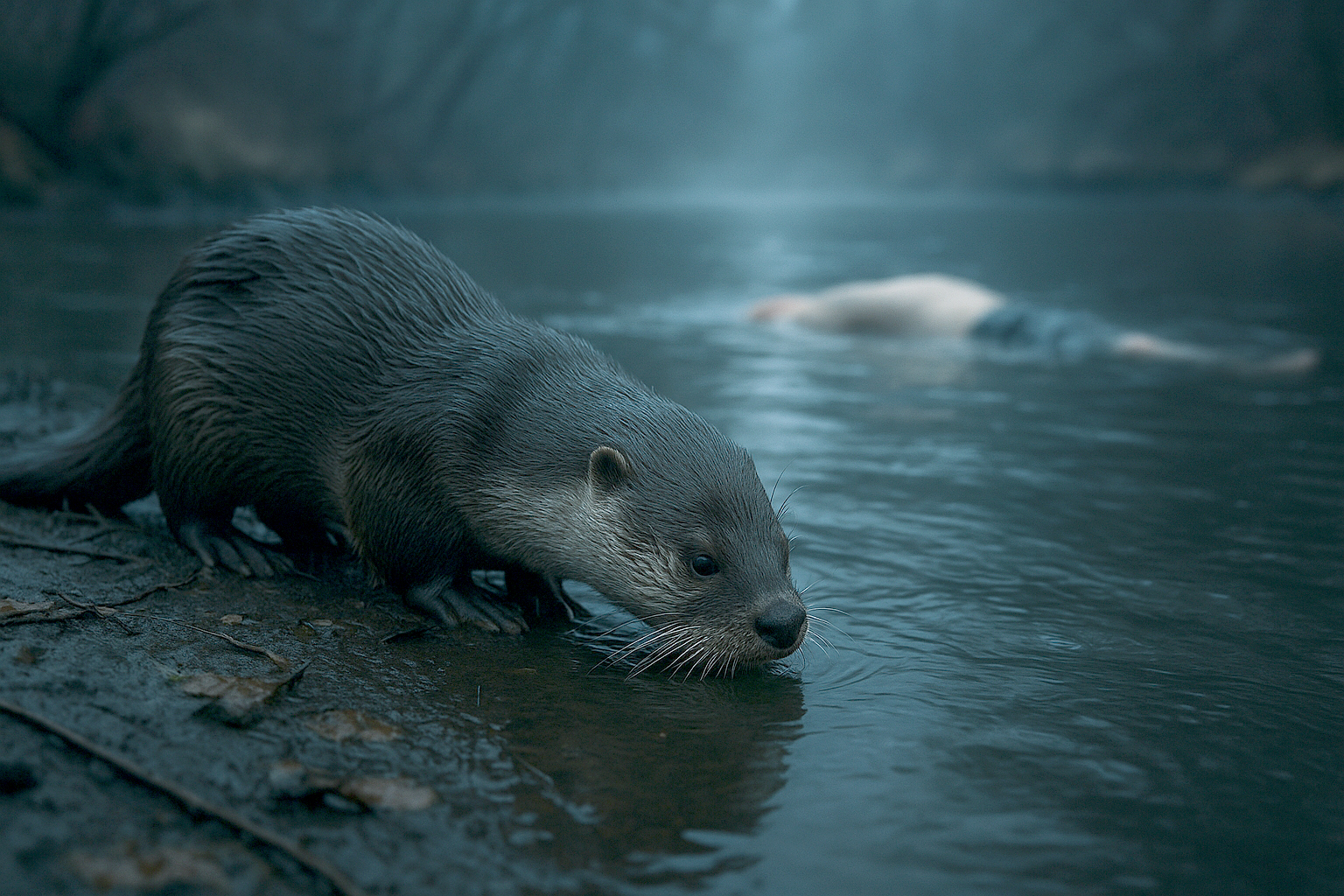
In the United States, an otter named Splash has been trained to detect submerged human bodies using its extraordinary sense of smell. This unprecedented forensic initiative opens new perspectives for locating bodies in rivers and streams. In France, where four bodies were recently discovered in the Seine River, such a protocol could inspire the river brigades of the National Police and the National Gendarmerie, thereby enhancing the efficiency of judicial investigations.

Animals in the service of forensic science

For decades, animals have played a major role in criminal investigations and forensic science (see odorology and scent identification). Cadaver detection dogs are now indispensable assets in forensic investigations: they detect the volatile compounds associated with human decomposition and assist in locating buried or concealed bodies. However, in aquatic environments, these methods face significant limitations—reduced visibility, strong currents, and variable depths. It was under these conditions that an innovative idea emerged in Florida: assigning this mission to an animal perfectly adapted to aquatic environments. The otter—agile, fast, and gifted with an exceptional sense of smell—proved to be the ideal candidate. The Peace River K9 Search & Rescue association thus launched a pioneering program by training Splash, an Asian small-clawed otter who has become the world’s first “investigative otter.”

A unique training protocol: air bubbles simulating human decomposition

Splash’s training follows a precise and rigorous protocol. In the trainer’s backyard, pools were installed to create a controlled environment. The water is infused with air bubbles containing volatile organic compounds similar to those released by a decomposing human body.

The otter’s mission is clear: to detect these air bubbles invisible to the human eye. When it recognizes the scent, it immediately alerts its trainer by tugging on the mask he is wearing. This signal—simple yet effective—confirms the presence of a “target.” The concept relies on a remarkable and still little-studied ability: the otter can literally “taste” the air bubbles underwater, chemically detecting specific markers within them. Where divers and dogs reach their limits, the otter excels thanks to its natural ability to navigate complex and opaque aquatic environments.

The otter in the service of criminal investigations

American forensic authorities are closely monitoring this project. The FBI and the Florida Department of Law Enforcement have already expressed interest in this method, which could accelerate searches during criminal investigations or natural disasters. The potential applications are numerous:

  • Locating victims of drownings, homicides, or flash floods in lakes, ponds, rivers, or streams.
  • Quickly identifying submerged bodies in areas with low visibility.
  • Complementing existing search resources (divers, sonar, detection dogs).

For investigators and magistrates, this time saving is crucial: the discovery of a submerged body can provide essential forensic evidence (signs of violence, medico-legal analyses) before further decomposition occurs, allowing the judicial investigation to progress more rapidly.

Could France take inspiration from Splash?

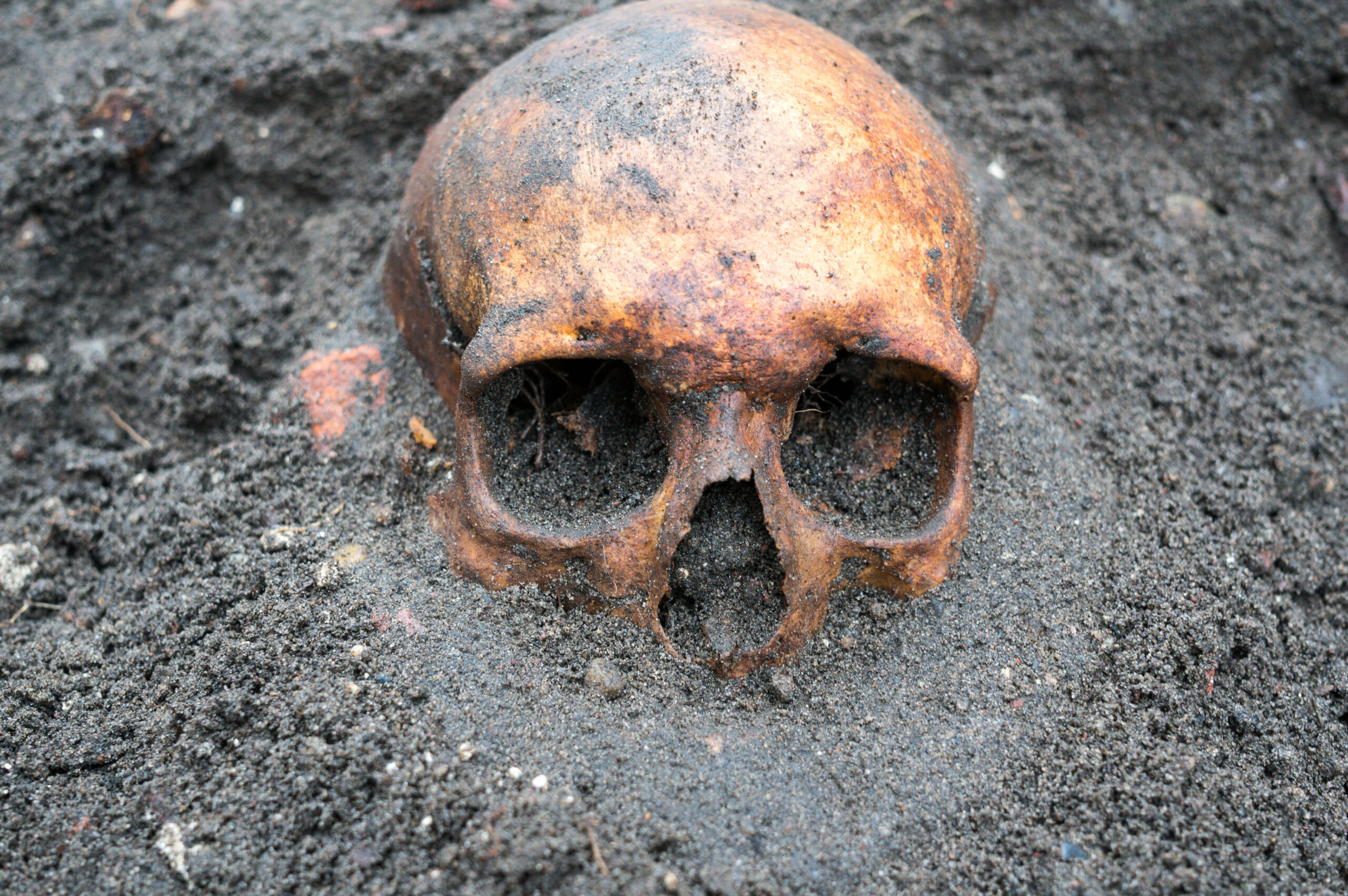
En France, la découverte de quatre corps dans la Seine In France, the discovery of four bodies in the Seine River on August 13, 2025, in Choisy-le-Roi (Val-de-Marne), served as a reminder of how complex the search for submerged bodies remains. The river brigades of the National Gendarmerie and the National Police already deploy divers, sonar equipment, and cadaver detection dogs to locate victims. Yet, despite these resources, some cases remain unsolved due to the absence of recovered bodies. The use of animals such as otters could provide a valuable complementary tool. Their acute sense of smell, agility, and ability to operate underwater could increase the likelihood of discoveries—particularly in rivers like the Seine, where visibility is almost zero and currents can carry bodies far from their original immersion point. Such a system could also prove useful in other environments: dams, canals, or large ponds. In homicide or missing person cases, any technology or protocol capable of accelerating the location of a body represents a major asset for judicial investigations.

Limitations and ethical considerations

While the method has generated interest, it also raises several important questions. Training otters requires time, specialized expertise, and impeccable ethics concerning animal welfare. Integrating such animals into official search systems would necessitate strict protocols, scientific validation, and an appropriate legal framework. However, as with cadaver detection dogs, the potential benefits are such that a gradual adoption of this approach does not seem unrealistic. Investigators dealing with sensitive cases—such as homicides or disappearances—know how decisive each additional tool can be.

Conclusion

The story of Splash illustrates a new synergy between nature and forensic science. Where technology and divers reach their limits, animals endowed with extraordinary senses remind us that forensic investigation can also draw upon the living world. While the idea of integrating otters into river brigade operations may seem unconventional, it nonetheless represents a credible prospect: enhancing the efficiency of investigations and improving the chances of swiftly locating submerged bodies.

Références :

  • IFLScience – Meet Splash, the world’s first search-and-rescue otter hunting for missing people in Florida, consultable ici.
  • Popular Science – This otter is training to be a search and rescue diver, consultable ici.
  • Interesting Engineering – US otter trained for underwater search and rescue, consultable ici.

Heartbeat Detection as an Anti-Deepfake Tool

Deepfake videos generated by artificial intelligence are becoming increasingly realistic, threatening the integrity of digital evidence. To address this challenge, Dutch researchers have developed an innovative method to detect deepfakes using a previously overlooked biological marker: the heartbeat. Still under scientific validation, this approach could become a valuable tool in digital forensic investigations.

A biological signal impossible to fake?

At the core of this innovation is a team from the Netherlands Forensic Institute (NFI), working with the University of Amsterdam. Their method relies on remote photoplethysmography (rPPG), a technique that detects subtle color variations in facial skin—on the forehead, around the eyes, or along the jawline—caused by blood flow at each heartbeat. Current deepfake algorithms are unable to simulate these micro-variations consistently, opening a promising path for detecting manipulated content.

An idea revived by technological progress

The concept dates back to 2012, when Professor Zeno Geradts explored video footage in criminal cases to assess whether the filmed individuals were alive. At the time, a MIT study had demonstrated that heart rate could be extracted from facial videos, but video compression destroyed the signal. Today, modern compression technologies preserve these micro-visual variations far better. The NFI team identified 79 facial points of interest to measure the signal and compared the results to biometric data from clinical sensors and smartwatches. Findings are encouraging, though some limitations remain—particularly with darker skin tones.

Figure 1. Principle of rPPG.
The absorption and reflection of light by the skin vary depending on hemodynamic activity under light sources (sunlight, lamps, etc.). These variations are recorded by imaging devices (cameras, webcams, smartphone lenses, etc.) as videos or images. Through algorithmic analysis, rPPG curves representing physiological information can be extracted from these videos.

A complementary tool for digital forensics

Heartbeat detection does not replace existing authentication methods but adds a valuable new dimension to forensic video examination. Other approaches remain crucial in the authentication process, such as analyzing electrical network frequency (ENF) traces embedded in images, identifying the recording sensor through its digital fingerprint (PRNU), or carrying out visual/automated checks of blinking patterns, abnormal movements, or generation artifacts (like a hand with six fingers). By combining these methods, experts can strengthen the reliability of their conclusions and stay ahead of forgers’ evolving tactics.

Robustness lies in combining traditional forensic techniques with AI-based approaches, rather than depending on one unique method.

A technological cat-and-mouse game

As new detection methods emerge, deepfake creators will inevitably attempt to circumvent them. In the near future, algorithms may try to artificially embed biological signals such as heartbeats into fake videos. This makes ongoing technological monitoring essential to stay one step ahead. As Geradts emphasizes, robustness lies in combining traditional forensic techniques with AI-based approaches, rather than depending on one unique method.

Towards judicial integration?

This approach is not yet deployed in real-world investigations—it is still undergoing scientific validation, with an academic publication expected in the coming months. However, researchers hope that in specific cases, particularly with high-quality videos, this method could soon be implemented. It opens a promising new avenue in the fight against digital evidence manipulation, leveraging a hard-to-fake truth: human physiology.

Références :

  • Geradts, Z., Pronk, P., & de Wit, S. (2025, mai). Heartbeat detection as a forensic tool against deepfakes. Présentation à l’European Academy of Forensic Science Conference (EAFS), Dublin.
  • Computer Weekly. (2025, 24 juillet). Dutch researchers use heartbeat detection to unmask deepfakes. Read here.
  • ForensicMag. (2025, 30 mai). Scientist Develops Method to Use Heartbeat to Reveal Deepfakes. Read here.
  • Amsterdam AI. (2025, 27 mai). Hartslaganalyse helpt deepfakes te ontmaskeren. Read here
  • DutchNews.nl. (2025, 25 mai). Dutch forensic experts develop deepfake video detector using heartbeat signals. Read here.
  • Poh, M.-Z., McDuff, D., & Picard, R. W. (2010). Advancements in non-contact, automated cardiac pulse measurements using video imaging. Massachusetts Institute of Technology (MIT) Media Lab.

Touch DNA: a new approach to better understand the traces left behind

In criminal investigations, DNA analysis plays a central role in identifying the perpetrators of crimes and offenses. However, not all biological traces provide the same type of information. Touch DNA—deposited involuntarily on a surface after simple contact—remains challenging to interpret for forensic experts.

Why do some individuals leave more DNA than others? A recent study conducted by researchers at Flinders University in Australia proposes an innovative method to objectively assess this variability. By examining the individual propensity to shed skin cells, the team opens new perspectives in forensic genetics and the interpretation of biological traces at crime scenes.

A genuine interindividual variability

Some individuals, described as “good shedders,” naturally deposit large quantities of skin cells on objects they handle. Others, by contrast, leave only minimal traces. This difference, long observed by forensic biologists, complicates the interpretation of DNA results, particularly when assessing the likelihood of direct contact between a person and an object.

Until now, reliably and reproducibly quantifying this variability has been difficult. The Australian study specifically addresses this gap, providing a rigorous scientific protocol.

A simple and reproducible measurement protocol

The researchers developed a protocol based on a series of controlled contacts carried out by 100 participants, each asked to touch a standardized surface. The deposited cells were then:

  • Stained with a fluorescent marker,
  • Counted using microscopy,
  • Subjected to genetic analysis to confirm the presence of recoverable DNA.

The results showed that, for 98 out of 100 participants, the level of cell deposition was stable and reproducible over time. This protocol allows individuals to be classified into three categories: high, moderate, or low skin cell shedders.

A tool to better contextualize touch DNA evidence

The value of this method extends beyond biology. It may serve as a tool for judicial contextualization. For instance, a suspect identified as a high shedder could account for the abundant presence of their DNA on an object without having taken part in the offense. Conversely, the absence of DNA from a low shedder does not exclude the possibility of contact.

This information could be incorporated into likelihood ratio calculations used in DNA interpretation, thereby strengthening the robustness of forensic assessments.

Future perspectives for forensic science

The proposed method has several advantages: it is inexpensive, easy to implement in the laboratory, and could be adapted to various objects and realistic conditions (different surfaces, durations of contact, humidity). Further validation studies are still required before widespread adoption. Ultimately, however, this approach could be integrated into routine biological trace analysis, providing valuable support to magistrates and investigators in evaluating the probative value of DNA evidence.

References

  • Petcharoen P., Nolan M., Kirkbride K.P., Linacre A. (2024). Shedding more light on shedders. Forensic Science International: Genetics, 72, 103065, read here.
  • Flinders University. (2024, August 22). Heavy skin shedders revealed: New forensic DNA test could boost crime scene investigations. ScienceDaily, read here.

Uncovering the meaning of suspicious injuries in cases of child abuse

There is a certain difficulty in objectively identifying a cigarette burn in a forensic context, particularly when the victim cannot testify. Such lesions are of particular relevance in cases of suspected child abuse. Until now, diagnoses have relied mainly on the morphological appearance of the injuries, with no standardized tool to support a conclusion based on material evidence.

A striking clinical case of child abuse

A team from the Laboratory of Histological Pathology and Forensic Microbiology at the University of Milan investigated a suspected case of child abuse that resulted in the death of a child. Three circular lesions suggestive of cigarette burns were found on the body. A cigarette butt collected nearby further supported the suspicion of an intentional act. The challenge was to determine whether these marks were the result of deliberate harm. However, visual inspection and even conventional histology cannot always confirm the exact origin of such lesions. Hence the value of turning to a more refined and objective method.

The SEM–EDX method: a microscopic zoom on the lesion

Scanning electron microscopy (SEM) allows the morphology of the injured skin to be observed with extreme precision, while energy-dispersive X-ray spectroscopy (EDX) identifies the chemical elements present on the surface of the lesions. This analysis relied on internal calibration, applied both to samples of injured skin and to cigarette fragments collected at the scene.

Elemental signatures of an intentional act

The results revealed a circular lesion with a reddish base, consistent with intense thermal contact. The chemical composition detected by EDX contained elements typically associated with tobacco combustion, in particular sulfur trioxide and phosphorus oxides, confirming combustion rather than mere environmental residues. Combined with the histological findings, this analysis demonstrated that the injury had occurred prior to death, providing an objective element supporting the likelihood of abuse.

A tool to strengthen forensic expertise

The study demonstrates that SEM–EDX analysis, combined with histology, represents a significant advancement in the characterization of suspicious lesions in the context of child abuse. It moves beyond visual assessment to provide objective and reproducible data, essential in judicial proceedings. By overcoming the limitations of visual inspection, this approach delivers results based on reproducible physico-chemical evidence, thereby reinforcing the robustness of forensic conclusions in light of judicial requirements.

Conclusion

This study paves the way for broader integration of analytical microscopy into forensic practices. By combining scientific rigor with judicial investigation, it offers a robust method for clarifying the nature of lesions whose origin often remains uncertain. The approach could also be applied to other types of injuries, such as those caused by heat sources or chemical agents. This progress deserves to be extended and validated on a larger number of cases in order to refine its reliability.

Références :

  • Tambuzzi S. et al. (2024). Pilot Application of SEM/EDX Analysis on Suspected Cigarette Burns in a Forensic Autopsy Case of Child Abuse. American Journal of Forensic Medicine & Pathology, 45(2), 135‑143. Read here.
  • Faller-Marquardt M., Pollak S., Schmidt U. (2008). Cigarette Burns in Forensic Medicine. Forensic Sci. Int., 176(2–3), 200–208
  • Maghin F. et al. (2018). Characterization With SEM/EDX of Microtraces From Ligature in Hanging. Am. J. Forensic Med. Pathol., 39(1), 1–7, read here.

How do nature indicates the presence of a corpse ?

What if fungal spores and pollen grains could reveal the secrets of clandestine graves? That is the hypothesis explored by an international team of researchers in Colombia, who conducted a pioneering experiment combining mycology and palynology in a forensic context. 

A biological approach to detecting illegal graves

In an experimental project carried out in Bogotá, two graves simulating clandestine burials were dug — one empty, the other containing a pig cadaver (a standard human body substitute in forensic science). Soil samples were collected and analyzed at different depths to study fungal and pollen communities composition. The aim of the study was to determine whether decomposed organic remains alter the soil’s microbial and plant-based communities, and whether these biological signatures could serve as spatial and temporal indicators in criminal investigations. 

Revealing fungal and pollen richness

The results showed that soil from the pits containing a carcass exhibits greater fungal richness (higher species diversity), notably with species such as Fusarium oxysporum and Paecilomyces, whose frequency increased in the presence of decomposition. These organisms, capable of degrading nitrogen-rich compounds such as keratin, could serve as indicators of buried organic remains.

Fungal structures of Fusarium oxysporum observed under optical microscopy.
A and B: macroconidia; C: chlamydospores. © David Esteban Duarte-Alvarado

On the palynology side, pollen grains identified at 50 cm depth—including Borago officinalis, Poa sp., and Croton sonderianus—are typical of the dry season. In contrast, the pollens found at 30 cm correspond to the rainy season. This stratified distribution could allow investigators to estimate the burial and exhumation periods with greater accuracy.

Integrating soil biology into criminal investigations

This study is the first to provide experimental data on mycology and palynology in an equatorial tropical context, a field largely unexplored in forensic science until now. It paves the way for a more systematic integration of these disciplines in crime scene investigations involving clandestine graves or the search for buried remains. While preliminary, the findings demonstrate the value of biological approaches as a complement to conventional forensic methods especially in regions where climatic conditions influence decomposition dynamics.

Conclusion

This study is part of a broader research effort into biological indicators left by buried bodies. After trees and roots that can signal underground anomalies, it is now fungi and pollen that emerge as silent witnesses of clandestine deaths. This microbiological approach expands the toolkit of forensic archaeology, as practiced by experts such as those from the French Gendarmerie. By combining invisible biological traces with conventional excavation and stratigraphic analysis techniques, it enables a more precise reading of the soil—and the criminal stories it may conceal.

Reference :
Tranchida, M. C., et al. (2025). Mycology and palynology: Preliminary results in a forensic experimental laboratory in Colombia, South America. Journal of Forensic Sciences.
Full article here.

When artificial intelligence reads the signs of death

Estimating the postmortem interval (PMI) largely relies on identifying (scoring) the stage of decomposition (SOD) of the body. Until now, this crucial step has been performed primarily by human experts using semi-objective visual methods. However, these approaches suffer from significant limitations: subjectivity, processing time, and difficulties in handling massive datasets.

A recent study conducted by the University of Tennessee investigates the contribution of artificial intelligence (AI) to automating this classification. Drawing on a dataset of over 1.5 million images of decomposing bodies documented under real conditions between 2011 and 2023, the researchers trained two convolutional neural network (CNN) models: Inception V3 and Xception.

A segmented anatomical approach based on deep learning

The study employed a strategy of decomposition stage scoring by anatomical region (head, trunk, and limbs), consistent with the methods of Megyesi (4 stages) and Gelderman (6 stages). Images were automatically sorted and then manually annotated by an expert according to these reference systems. The AI models were subsequently trained through transfer learning and tested on unseen images.

Performance results are highly promising, particularly with the Xception model, which achieved a high F1-score for both methods—an indicator of an AI model’s ability to generate predictions that are both accurate and comprehensive. Results were more modest for the limbs, owing to variability in photographic conditions.

A reliability equivalent to human experts?

To evaluate the performance of artificial intelligence against human experts, the researchers conducted an inter-rater test on 300 thoracic images. Three specialists classified the decomposition stages of these images using the two recognized methods, and their results were compared with those generated by the AI.

Agreement was assessed using Fleiss’ Kappa coefficient. For the Megyesi method, results revealed a “substantial” agreement between AI classifications and those of human experts (κ = 0.637), a score very close to that observed among the experts themselves (κ = 0.67). These findings highlight the significant alignment of AI with expert evaluations, thereby reinforcing the validity and relevance of this automated approach.

Challenges to overcome for operational integration

Annotation carried out by a single expert introduces bias, while the use of a unique environmental context limits the generalizability of the results. Lower performance on limb regions highlights the need for greater data diversification, particularly through the inclusion of varied climatic conditions. A multicenter dataset annotated by multiple experts would provide a more robust reference base, ensuring improved generalization and increased reliability of the models.

Perspectives: toward AI-augmented forensics

This study represents a step forward in the automation of taphonomic analysis. Other work, such as that of Smith et al. (2024) using Bayesian models, or the growing use of 3D imaging and the necrobiome, suggest a convergence of AI, biological, and environmental approaches for a more accurate and less subjective estimation of the PMI.

Automating the assessment of decomposition stage allows for substantial time savings while reducing inter-observer variability. However, further efforts are needed to expand datasets and to develop standardized annotation protocols. The integration of algorithms such as those described here could transform forensic practice by facilitating the exploitation and analysis of large image databases, as well as their application in crisis situations (disasters, conflicts).

References :

  • Nau, A.-M. et al. (2024). Towards Automation of Human Stage of Decay Identification: An Artificial Intelligence Approach. arXiv:2408.10414.
  • Megyesi, M.S. et al. (2005). Using accumulated degree-days to estimate the postmortem interval from decomposed human remains. Journal of Forensic Sciences, 50(3), 618–626.
  • Gelderman, H. et al. (2018). The development of a post-mortem interval estimation for human remains found on land in the Netherlands. Int. J. Legal Med., 132(3), 863–873.
  • Smith, D.H. et al. (2024). Modeling human decomposition: a Bayesian approach. arXiv:2411.09802.
  • Infante, D. (2025). How AI and 3D Imaging are Transforming Body Farm Research. AZoLifeSciences.
  • Piraianu, A.-I. et al. (2023). Enhancing the evidence with algorithms: how artificial intelligence is transforming forensic medicine. Diagnostics, 13(18), 2992.

When teeth talk : How dental tartar serves toxicology

Initially exploited in archaeology, dental calculus is now revealing its potential in forensic science. It retains traces of ingested substances, opening the way to post-mortem analysis of drug intake and psychoactive compounds.

Dental calculus: A neglected but valuable matrix

Dental calculus forms through the gradual mineralization of dental plaque, a biofilm composed of saliva, microorganisms, and food residues. This process traps various compounds present in the oral cavity, including xenobiotics such as drugs or their metabolites. Its crystalline structure grants this matrix an excellent preservation properties for the substances it contains, while making it resistant to external degradation, including in post-mortem or archaeological contexts.

A new path for tracking illicit substances

Recently, a research team demonstrated the feasibility of a toxicological approach based on the analysis of dental calculus using liquid chromatography coupled with tandem mass spectrometry (LC-MS/MS). In a study involving ten forensic cases, the researchers detected 131 substances in tartar, compared to 117 in blood—sometimes in higher concentrations within the tartar. The method enabled the identification of common drugs such as cocaine, heroin, and cannabinoids, even in cases where they were no longer detectable in conventional matrices (Sørensen et al., 2021). These substances, absent from the blood, were often present in higher concentrations in dental tartar.

A long-lasting and discreet witness

This approach offers several clear advantages. It allows the detection of substance use weeks or even months after ingestion. Tartar sampling is non-invasive and applicable to skeletal remains, making it particularly relevant in archaeological and forensic anthropology contexts. It can help reconstruct consumption habits, medical treatments, or causes of death in situations where blood, urine, or hair are unavailable.

A promising method to be further developed

One of the main strengths of this technique lies in its ability to exploit a matrix that is often overlooked but commonly available on teeth. Only a few milligrams are needed to conduct a reliable analysis—provided the trapped substances remain stable over time. This method also opens the possibility of broadening the range of detectable compounds, pending further validation.

While promising, this avenue still requires additional research to standardize protocols, assess the long-term stability of molecules, and fully integrate this approach into routine forensic toxicology practices. Although still in its exploratory phase, the method offers remarkable potential for the use of alternative matrices and opens new perspectives for forensic toxicology.

Reference:

  • Sørensen LK, Hasselstrøm JB, Larsen LS, et al. Entrapment of drugs in dental calculus: detection validation based on test results from post-mortem investigations. Forensic Sci Int 2021; 319: 110647.
  • Reymond C, Le Masle A, Colas C, et al. A rational strategy based on experimental designs to optimize parameters of a liquid chromatography-mass spectrometry analysis of complex matrices. Talanta 2019; 205: 120063.
  • Radini A, Nikita E, Buckley S, Copeland L, Hardy K. Beyond food: The multiple pathways for inclusion of materials into ancient dental calculus. Am J Phys Anthropol 2017; 162: 71–83.
  • Henry AG, Piperno DR. Using plant microfossils from dental calculus to recover human diet: a case study from Tell al-Raqā’i, Syria. J Archaeol Sci 2008; 35: 1943–1950.

Bedbugs: a new weapon for forensic science?

Malaysian researchers have explored the potential of tropical bedbugs, Cimex hemipterus, as a new source of human DNA in forensic investigations. Typically overlooked in crime scene analyses due to the absence of visible traces, these insects may nevertheless carry, within their digestive tract, the DNA of the last human host they fed on. The study aimed to determine whether—and for how long—a usable human DNA profile could be extracted from the blood meal content of bedbugs, focusing on two key forensic genetic markers: STRs (Short Tandem Repeats) and SNPs (Single Nucleotide Polymorphisms).

Methodology and results

Laboratory-reared bedbug colonies were fed on human volunteers and subsequently sacrificed at different intervals (0, 5, 14, 30, and 45 days after feeding). DNA was extracted and subjected to STR and SNP analyses following standard forensic protocols. The results were conclusive: complete STR and SNP profiles could only be obtained on the day of feeding (day 0), while partial, though still informative, profiles remained detectable up to 45 days post-feeding. The SNP data were interpreted using the HIrisPlex-S system, allowing phenotype predictions (eye, skin, and hair colour) even from partial genetic information. Moreover, field-collected bedbugs confirmed the feasibility of STR profiling, occasionally revealing mixed DNA profiles—potentially indicating feeding from multiple human hosts.

These results open up a new avenue for forensic science: when traditional biological traces have disappeared or been cleaned away, bedbugs could remain at the scene and serve as reliable micro-reservoirs of human DNA, enabling investigators to identify individuals who were present or to establish a timeline of movements. However, several limitations must be taken into account. First, the analyses are time-consuming and require a rigorous protocol. The DNA profile becomes partial after a few days, and some loci are no longer detectable. Moreover, when an insect has fed on multiple individuals, mixed genetic signals can occur, making interpretation more complex.

The authors emphasize the need to validate these findings on a broader range of samples, including more human donors and various commercial STR/SNP kits. Controlled in situ tests on simulated crime scenes would also be useful to confirm the robustness of the method—particularly in comparison with other insects or biological intermediaries considered in forensic entomology.

Conclusion

In summary, this study demonstrates that human DNA preserved in the stomach of tropical bedbugs can be exploited for up to 45 days after feeding through STR and SNP analysis. Although a complete genetic profile can only be obtained immediately after feeding, these insects represent an innovative and promising resource for forensic science, especially in situations where conventional methods fail. Nevertheless, the approach requires strict protocols, further validation studies, and realistic crime-scene modelling before it can be used in judicial proceedings. Additional research will determine how this strategy can be integrated into the growing toolkit of forensic investigators and scientists.

Sources :

  • Kamal, M. M. et al. (2023)Human profiling from STR and SNP analysis of tropical bed bug (Cimex hemipterus) for forensic science, Scientific Reports, 13(1), 1173.
  • Chaitanya, L. et al. (2018)HIrisPlex-S system for eye, hair and skin colour prediction from DNA, Forensic Science International: Genetics, 35, 123–134.
  • Asia News Network (2023)Malaysian scientists discover bed bugs can play role in forensic investigations, Read full article.
  • ResearchGate – Publication originaleHuman profiling from STR and SNP analysis of tropical bed bug Cimex hemipterus for forensic science, Read full article.

Photogrammetry, Lasergrammetry, and Artificial Intelligence: A Technological Revolution

Forensics and emergency response are currently at a turning point with the growing integration of advanced technologies such as photogrammetry, lasergrammetry (LiDAR), and artificial intelligence (AI). These technologies not only provide unprecedented levels of accuracy and efficiency but also open up new avenues for investigation and intervention, profoundly reshaping traditional methodologies.

Photogrammétrie et Lasergrammétrie : des outils de précision

As a surveying expert and officer specializing in the drone unit of the Haute-Savoie Fire and Rescue Department (SDIS74), I have directly observed how these tools enhance the accuracy of topographic surveys and facilitate the rapid analysis of complex scenes. Photogrammetry enables 3D reconstruction of various environments using aerial images captured by drones equipped with high-resolution cameras. This process quickly generates detailed digital terrain models, which are critical in urgent or forensic interventions where every detail matters.

Road survey using photogrammetric methods, in true color. Credit: Arnaud STEPHAN – LATITUDE DRONE

It is possible to achieve extremely high levels of detail, allowing, for example, the identification of footprints by the depth left in the ground.

LiDAR scanning effectively complements photogrammetry by providing millimetric precision through the emission of laser beams that scan and model the environment in three dimensions. This technology is particularly effective in complex contexts such as dense wooded areas, steep cliffs, or rugged mountain terrain, where photogrammetry may sometimes struggle to capture all the necessary details.

To be more precise, LiDAR generally produces more noise on bare ground and hard surfaces compared to photogrammetry, which remains the preferred tool in such cases. However, in wooded areas, LiDAR can occasionally penetrate through to the ground and thus provide crucial information about the terrain, where photogrammetry may fail.

Photogrammetry only works during daylight, since it relies on photographic data in the visible spectrum.

Depending on the chosen flight altitudes and the type of sensor used, it is possible to achieve extremely high levels of detail, allowing, for example, the identification of footprints by the depth left in the ground. These technologies are already being used to precisely capture crime scenes. Traditionally, static scanners were used for this purpose, but drones now make it possible to greatly expand the capture perimeter while ensuring faster processing. This speed is crucial, as it is often imperative to capture the scene quickly before any change in weather conditions.

However, it is important to note that photogrammetry only works during daylight, since it relies on photographic data in the visible spectrum.

Topographic survey using LiDAR method and colored according to altitude. Vegetation differentiated in green. Credit: Arnaud STEPHAN – LATITUDE DRONE

Artificial Intelligence: towards automated and efficient analysis

The true revolution lies in the integration of these geospatial surveys into intelligent systems capable of massively analyzing visual data with speed and precision. In this regard, the OPEN RESCUE project, developed by ODAS Solutions in partnership with SDIS74 and the Université Savoie Mont-Blanc, stands as an exemplary case. This AI is fueled by an exceptional dataset of nearly 1.35 million images collected using various types of drones (DJI Mavic 3, DJI Matrice 300, Phantom 4 PRO RTK, etc.) across a remarkable diversity of environments, covering all seasons.

Illustration of OPEN RESCUE’s capabilities: a person isolated in the mountains during winter. Credit: Arnaud STEPHAN – ODAS SOLUTIONS

The robustness of the OPEN RESCUE AI is demonstrated by a maximum F1-score of 93.6%, a remarkable result validated through real field operations. The F1-score is a statistical indicator used to measure the accuracy of an artificial intelligence system: it combines precision (the number of correctly identified elements among all detections) and recall (the number of correctly identified elements among all those actually present). A high score therefore means that the AI effectively detects a large number of relevant elements while avoiding false detections. This intelligent system is capable of accurately detecting individuals as well as indirect signs of human presence such as abandoned clothing, immobilized vehicles, or personal belongings, thereby providing valuable and immediate assistance to rescue teams.

Collection of OPEN RESCUE training data with SDIS74 firefighters – Credit: Arnaud STEPHAN – ODAS SOLUTIONS

The arrival of this technology is radically transforming the way teams conduct their searches: it is now possible to methodically and extensively sweep entire areas, while ensuring that no relevant element has been missed by the AI in these zones. Although this does not replace canine units or other traditional methods, artificial intelligence provides a new and complementary level of thoroughness in the search process.

The arrival of this technology is radically transforming the way teams conduct their searches.

Practical Applications and Operational Results

In the field, the effectiveness of these technologies has been widely demonstrated. The autonomous drones used by our unit can efficiently cover up to 100 hectares in about 25 minutes, with image processing carried out almost in real time by OPEN RESCUE. This enables an extremely rapid response, ensuring optimal management of critical time during emergency interventions and missing-person searches.

Furthermore, the ability to precisely document the areas covered during operations provides a significant advantage in judicial contexts. The possibility of using these accurate 3D models and automatically analyzed data as evidence before courts offers greater transparency in judicial procedures and greatly facilitates the work of judges, investigators, and lawyers.

DJI Matrice 300 drone flying in a mountainous area – Credit: Arnaud STEPHAN – LATITUDE DRONE

Operational constraints and regulatory framework

The operational use of drones and these advanced technologies is subject to several strict regulatory constraints, particularly in terms of flight authorizations, privacy protection, data management, and air safety. In France, drones are regulated by the Direction Générale de l’Aviation Civile (DGAC – French Civil Aviation Authority), which imposes specific flight scenarios and precise protocols to be followed during missions.

In addition, the technical constraints of operations include the need for trained and regularly certified pilots, capable of carrying out missions safely and efficiently. Finally, roughly every six months, new innovative equipment is released, constantly bringing significant improvements such as higher capture speeds, better optical and thermal sensors, and the miniaturization of onboard LiDAR systems.

Conclusion

Ultimately, the growing integration of advanced technologies represents a decisive breakthrough in forensic sciences and emergency interventions, despite the operational and regulatory constraints to be taken into account. Their practical application not only enhances the efficiency and speed of operations but also opens up new possibilities for judicial analysis, thereby confirming their essential role in public safety and modern justice.

Entomotoxicology: the role of insects in forensic investigation

When the body no longer speaks, insects tell the truth. At the crossroads of toxicology and entomology, entomotoxicology turns these small organisms into key witnesses capable of revealing what time seeks to erase.

Insects serving the truth

In certain deaths, especially those linked to drug abuse or suicide, the victim’s body may remain undiscovered for several days or even months. Decomposition begins immediately after death, releasing gases and fluids that produce putrefaction odors. These effluents quickly attract insects such as flies, which lay their eggs in the body’s natural openings. The emerging larvae feed on the decomposing flesh, thereby accelerating the breakdown process [Forensic Entomology – Damien Charabidze].

As insect colonization progresses, it further accelerates the gradual decomposition of body tissues. This degradation also alters organic fluids and tissues such as urine, blood, and the liver, rendering traditional matrices used in forensic toxicological analyses unavailable or unreliable. At this stage, insects—particularly blowfly larvae—become especially valuable for toxicological examination.

Discovery of larvae on a decomposing body

Because of their abundance, ease of collection, and resistance to environmental conditions, necrophagous insect larvae can be sampled from various regions of the body where they are present. This choice is critical, as the ante- and postmortem redistribution of substances (medications, drugs, toxins) within the body may vary between tissues, leading to qualitative and quantitative differences in the insects. Collecting multiple samples from different area therefore improves the accuracy and reliability of qualitative results. Numerous studies have demonstrated their potential to reveal toxic substances where traditional methods have failed [1–4]. To date, entomotoxicological analyses allow only for the qualitative detection of toxic substances present in the decedent’s body. This means that one can confirm the presence or absence of a drug, poison, or medication in the tissues consumed by the larvae, but not yet reliably determine its concentration.

Such results can therefore only support a hypothesis of intoxication. However, establishing whether the detected quantity was lethal is not yet possible, as this would require a more reliable quantitative approach.

In addition, the toxicologist must bear in mind that these small organisms are capable of metabolizing substances and producing metabolites similar to those generated by the human body, despite the usual complexity of such biotransformations in humans. Research into this phenomenon is still in its early stages.

Forensic entomological methods can assist in determining the minimum time elapsed between death and the discovery of the body.

Drugged insects, misleading dating!

In a criminal investigation, a key factor to consider—especially in cases involving decomposed bodies—is the minimum postmortem interval (minPMI). This refers to the time elapsed between the moment when the first insects colonized the body and its discovery (Fig. 1).

Fig. 1. Simplified diagram illustrating the difference between PMI and minPMI.

It is referred to as a minimum because this estimate does not begin at the exact time of death but at the moment of the first insect colonization, which occurs shortly after death, ranging from a few minutes to several hours depending on environmental factors. Once rigor mortis, livor mortis, and the cooling of the body to ambient temperature have passed, it becomes increasingly difficult to estimate the time elapsed since death. The body then enters the putrefaction phase, during which forensic entomological methods can assist in determining the minimum time elapsed between death and the discovery of the body.

However, if the victim had consumed drugs prior to death, this can significantly affect the development of necrophagous insects by accelerating or delaying their growth.

Forensic entomologists measure the size and study the developmental stage of the larvae present on the body, and by taking into account factors such as ambient temperature, the insect species identified, and data on necrophagous species succession [5, 6], they compare these findings with the life cycle of the insects concerned (Fig. 2).

Fig. 2. Life cycle of necrophagous flies (Diptera).

Since certain species colonize a cadaver very soon after death, they make it possible to determine the day of initial colonization and thereby estimate a minimum postmortem interval (minPMI).

However, if the victim had consumed drugs prior to death, this can significantly affect the development of necrophagous insects by accelerating or delaying their growth. Comparison with their life cycle is therefore biased, leading to overestimation or underestimation of the minPMI. This is why studies have been conducted to evaluate the impact of specific drugs on insect development, with the aim of eventually incorporating these variability ranges into more accurate minPMI estimations [7–12]. The way in which insects metabolize or fail to metabolize ingested toxic substances remains uncertain.

Limitations of entomotoxicology

Although insects can provide valuable information, entomotoxicology is not without limitations.

First, the environment plays a critical role: insect development is highly dependent on temperature, humidity, and climate. If the body is, for example, exposed to extreme heat or strong winds, insect colonization may differ. Ambient temperature directly influences insect survival and development. Harsh environmental conditions may kill the insects or slow their growth, thereby biasing or preventing analysis.

Moreover, beyond development, insect colonization itself can also be disrupted. For instance, when a body is submerged in water or covered (by clothing, tarpaulins, soil, debris, etc.), insect access is hindered. This may alter the dynamics of colonization and consequently the way insects feed on the body.

Another challenge in entomotoxicology is the uncertainty regarding how insects metabolize or fail to metabolize toxic substances they ingest. Unlike humans, their metabolism and the ways in which they store or eliminate toxic substances are poorly understood. As a result, it is difficult to establish a correlation between the amount of toxic substances detected in insects and the dose ingested by the deceased, making it problematic to confirm a lethal dose in the body and therefore to substantiate and accept a prior hypothesis of fatal intoxication.

Furthermore, methods for extracting, purifying, and analyzing toxic substances in insect larval tissues are not yet standardized. Each study must be adapted according to the substances being investigated.

Conclusion

Forensic entomotoxicology thus illustrates how insects could in the future become key witnesses in the resolution of criminal investigations. It opens up promising perspectives for forensic medicine by broadening the range of possibilities when no other biological sample is available. With advances in research, entomotoxicology may become an even more precise tool—not only qualitative but also quantitative—and essential in contributing to the establishment of truth in real forensic casework.

Bibliographie :

  • [1] Campobasso, C., Gherardi, M., Caligara, M., Sironi, L., & Introna, F. (2004c). Drug analysis in blowfly larvae and in human tissues : a comparative study. International Journal Of Legal Medicine, 118(4). https://doi.org/10.1007/s00414-004-0448-1
  • [2] Groth, O., Franz, S., Fels, H., Krueger, J., Roider, G., Dame, T., Musshoff, F., & Graw, M. (2021). Unexpected results found in larvae samples from two postmortem forensic cases. Forensic Toxicology, 40(1), 144‑155. https://doi.org/10.1007/s11419-021-00601-x
  • [3] Levine, B., Golle, M., & Smialek, J. E. (2000b). An unusual drug death involving maggots. American Journal Of Forensic Medicine & Pathology, 21(1), 59‑61. https://doi.org/10.1097/00000433-200003000-00010
  • [4] Beyer, J., Enos, W., & Stajić, M. (1980b). Drug Identification Through Analysis of Maggots. Journal Of Forensic Sciences, 25(2), 411‑412. https://doi.org/10.1520/jfs12147j
  • [5] Lutz, L., Verhoff, M. A., & Amendt, J. (2018). Environmental factors influencing flight activity of forensically important female blow flies in Central Europe. International Journal Of Legal Medicine, 133(4), 1267 1278. https://doi.org/10.1007/s00414-018-1967-5
  • [6] Matuszewski, S. (2021). Post-Mortem Interval Estimation Based on Insect Evidence : Current Challenges. Insects, 12(4), 314. https://doi.org/10.3390/insects12040314
  • [7] Boulkenafet, F., Dob, Y., Karroui, R., Al-Khalifa, M., Boumrah, Y., Toumi, M., & Mashaly, A. (2020). Detection of benzodiazepines in decomposing rabbit tissues and certain necrophagic dipteran species of forensic importance. Saudi Journal Of Biological Sciences, 27(7), 1691‑1698. https://doi.org/10.1016/j.sjbs.2020.04.044
  • [8] Gosselin, M., Di Fazio, V., Wille, S. M., Del Mar Ramírez Fernandez, M., Samyn, N., Bourel, B., & Rasmont, P. (2011b). Methadone determination in puparia and its effect on the development of Lucilia sericata (Diptera, Calliphoridae). Forensic Science International, 209(1‑3), 154‑159. https://doi.org/10.1016/j.forsciint.2011.01.020
  • [9] El-Samad, L. M., El-Moaty, Z. A., & Makemer, H. M. (2011). Effects of Tramadol on the Development of Lucilia sericata (Diptera : Calliphoridae) and Detection of the Drug Concentration in Postmortem Rabbit Tissues and Larvae. Journal Of Entomology, 8(4), 353‑364. https://doi.org/10.3923/je.2011.353.364
  • [10] Bourel, B., Hédouin, V., Martin-Bouyer, L., Bécart, A., Tournel, G., Deveaux, M., & Gosset, D. (1999). Effects of Morphine in Decomposing Bodies on the Development of Lucilia sericata (Diptera : Calliphoridae). Journal Of Forensic Sciences, 44(2), 354‑358. https://doi.org/10.1520/jfs14463j
  • [11] Zou, Y., Huang, M., Huang, R., Wu, X., You, Z., Lin, J., Huang, X., Qiu, X., & Zhang, S. (2013). Effect of ketamine on the development of Lucilia sericata (Meigen) (Diptera : Calliphoridae) and preliminary pathological observation of larvae. Forensic Science International, 226(1‑3), 273‑281. https://doi.org/10.1016/j.forsciint.2013.01.042
  • [12] O’Brien, C., & Turner, B. (2004). Impact of paracetamol on Calliphora vicina larval development. International Journal Of Legal Medicine, 118(4), 188‑189. https://doi.org/10.1007/s00414-004-0440-9