How can an internal lesion go unnoticed during autopsy yet may have potentially caused death? In forensic medicine, understanding internal trauma is essential to reconstructing the sequence of a violent event. Among such injuries, those involving the vertebral artery present a major challenge. Subtle and often concealed by bone structures, they frequently escape traditional examination methods. A recent technological breakthrough in forensic imaging offers a promising approach: combining fluoroscopy and micro-computed tomography (micro-CT) to analyze post-mortem vascular injuries with unprecedented precision.
Key artery, difficult access
The vertebral artery supplies vital regions of the nervous system, including the brainstem, cerebellum, and posterior areas of the brain. Even a minor injury can trigger a stroke, a rapid neurological collapse, or sudden death. Its anatomical pathway, deeply embedded within the cervical spine, makes it particularly difficult to explore. In a forensic context, a lesion affecting this artery represents a critical clue when analyzing a penetrating neck wound, often revealing a potentially lethal intent.
Forensic imaging to observe real-time blood flow
The vertebral artery supplies vital regions of the nervous system, including the brainstem, cerebellum, and posterior areas of the brain. Even a minor injury can trigger a stroke, a rapid neurological collapse, or sudden death. Its anatomical pathway, deeply embedded within the cervical spine, makes it particularly difficult to explore. In a forensic context, a lesion affecting this artery represents a critical clue when analyzing a penetrating neck wound, often revealing a potentially lethal intent.
Micro-CT: diving into the heart of the lesion
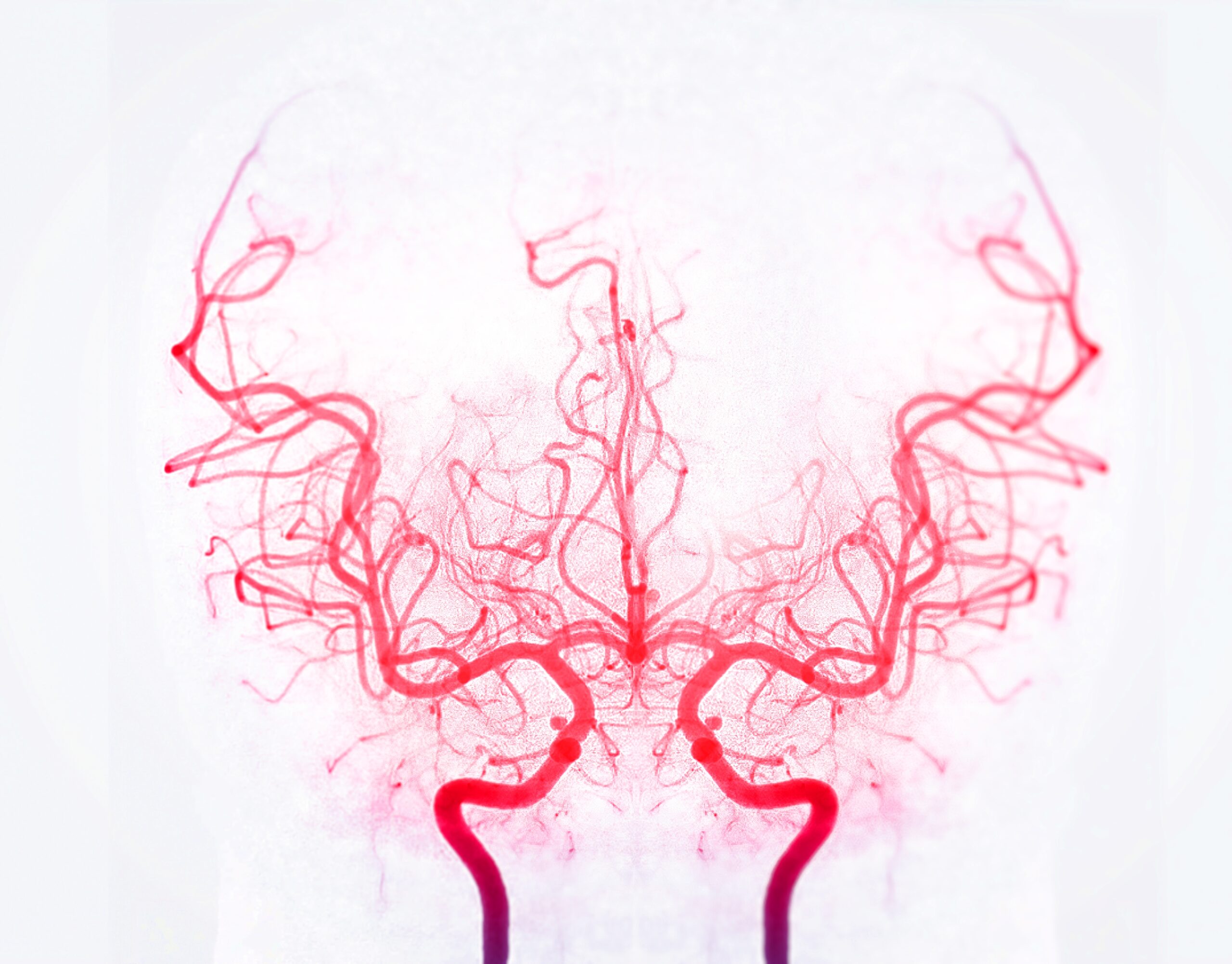
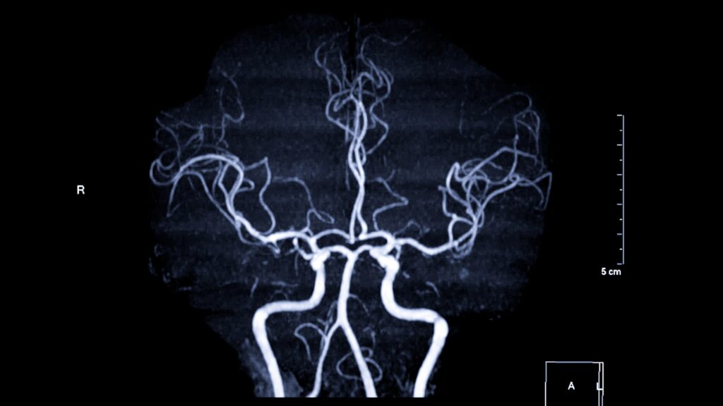
To overcome this limitation, researchers have turned to micro-computed tomography (micro-CT), a very high-resolution imaging technique. The sample is rotated during the acquisition of thousands of radiographic images, which are then reconstructed into a digital 3D model. This process reveals otherwise invisible details such as arterial wall tears, thrombi, dissections, or partial ruptures. These reconstructions allow for virtual dissections from multiple angles without altering the body, ensuring a high level of reproducibility, an invaluable feature in forensic investigations.
A standardized method serving both justice and medicine
The protocol developed by Secco and colleagues relies on ex situ imaging, meaning that the examination is performed on an artery extracted from the body. This approach overcomes several obstacles, such as advanced decomposition, previous surgery, complex trauma, or movement artifacts. With the injection of a contrast agent, the vascular network becomes clearly visualized, allowing for precise and stable documentation. These high-quality images serve as robust evidence admissible in court and represent a valuable resource for medical teams involved in planning neurosurgical or trauma-related procedures.
An educational and scientific tool
Beyond their diagnostic value, 3D reconstructions and fluoroscopic videos serve as outstanding educational tools. They allow for a strikingly realistic visualization of injury mechanisms and a deeper understanding of the biomechanics of penetrating trauma. This refined comprehension of the forces at play helps not only researchers characterize vascular lesions, but also engineers design more effective protective equipment and forensic experts accurately reconstruct the circumstances surrounding a violent act.
Towards a new standard in forensic medicine
Born from close collaboration between radiologists, pathologists, engineers, and chemists, this imaging protocol represents a major step forward in forensic practice. The growing accessibility of micro-CT equipment suggests its forthcoming integration into routine autopsies. With the continuous improvement of imaging technologies in terms of resolution, speed, and multi-contrast capacity, the prospect of non-invasive post-mortem vascular examinations is becoming increasingly realistic. In the long term, this method could be extended to other arterial regions (carotid, subclavian, intracranial), thereby deepening our overall understanding of vascular trauma.
Conclusion
At the crossroads of technology and forensic science, this approach combines precision, rigor, and innovation. By providing a three-dimensional and reproducible reading of internal injuries, it transforms the way stab wounds involving the vertebral artery are analyzed. This is a major advancement, serving both judicial truth and scientific knowledge, and it paves the way for a new generation of autopsies that are finer, more reliable, and better documented.
References :
Bioengineer.org. (2024). Detecting Vertebral Artery Stab Wounds with Imaging. Read here.
Secco, L., Franchetti, G., Viel, G. et al. Ex-situ identification of vertebral artery injuries from stab wounds through contrast-enhanced fluoroscopy and micro-CT. Int J Legal Med (2025). Read here.
Medscape. (2024). Vertebral Artery Anatomy. Read here.