Monthly Archives: July 2025

Heartbeat Detection as an Anti-Deepfake Tool

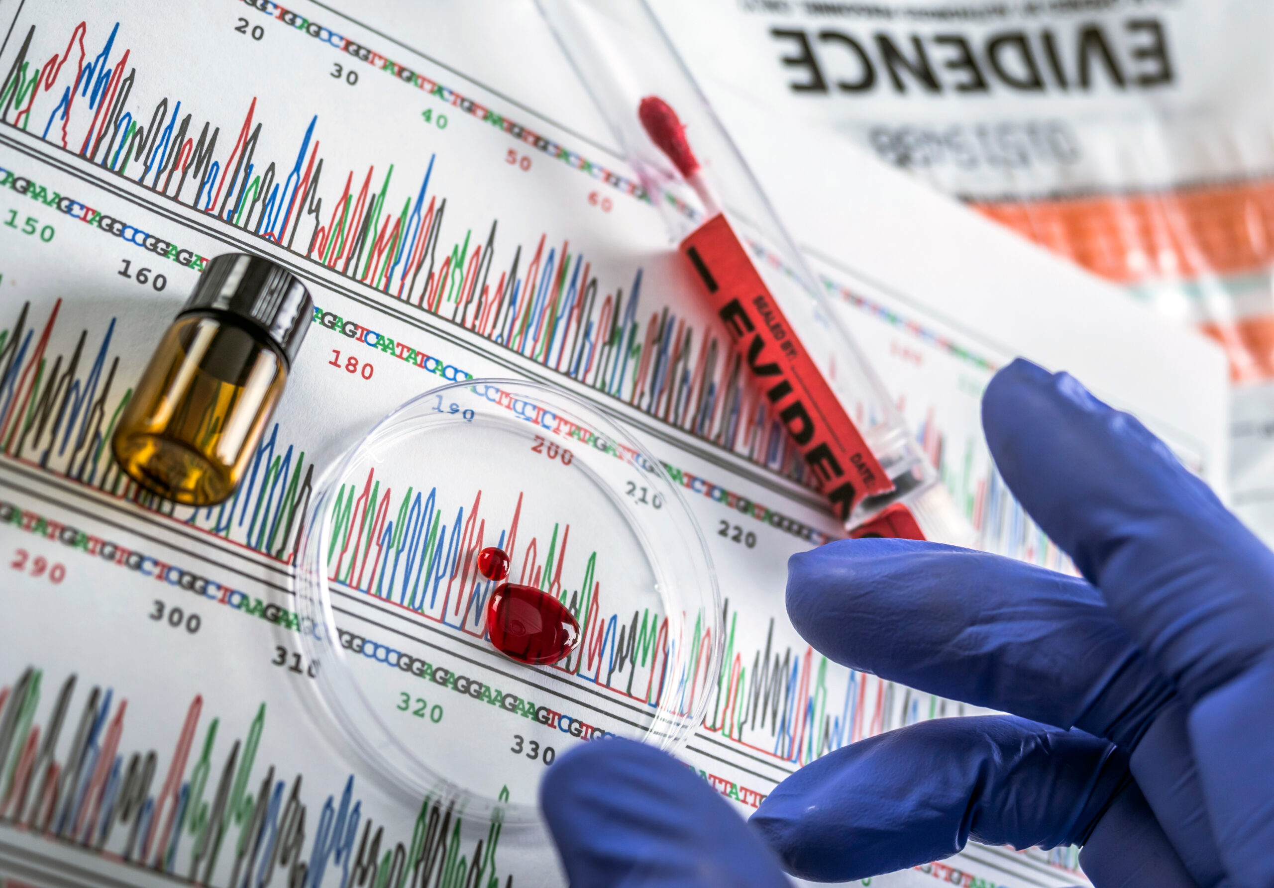
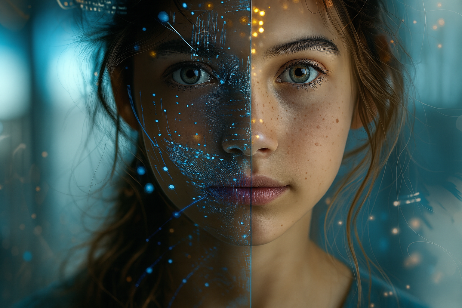
Deepfake videos generated by artificial intelligence are becoming increasingly realistic, threatening the integrity of digital evidence. To address this challenge, Dutch researchers have developed an innovative method to detect deepfakes using a previously overlooked biological marker: the heartbeat. Still under scientific validation, this approach could become a valuable tool in digital forensic investigations.

A biological signal impossible to fake?

At the core of this innovation is a team from the Netherlands Forensic Institute (NFI), working with the University of Amsterdam. Their method relies on remote photoplethysmography (rPPG), a technique that detects subtle color variations in facial skin—on the forehead, around the eyes, or along the jawline—caused by blood flow at each heartbeat. Current deepfake algorithms are unable to simulate these micro-variations consistently, opening a promising path for detecting manipulated content.

An idea revived by technological progress

The concept dates back to 2012, when Professor Zeno Geradts explored video footage in criminal cases to assess whether the filmed individuals were alive. At the time, a MIT study had demonstrated that heart rate could be extracted from facial videos, but video compression destroyed the signal. Today, modern compression technologies preserve these micro-visual variations far better. The NFI team identified 79 facial points of interest to measure the signal and compared the results to biometric data from clinical sensors and smartwatches. Findings are encouraging, though some limitations remain—particularly with darker skin tones.

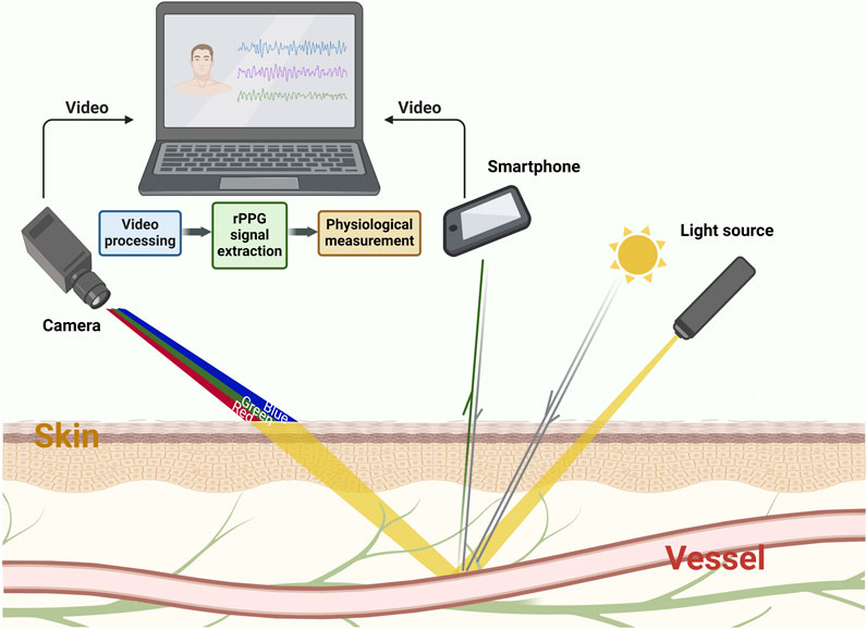
Figure 1. Principle of rPPG.
The absorption and reflection of light by the skin vary depending on hemodynamic activity under light sources (sunlight, lamps, etc.). These variations are recorded by imaging devices (cameras, webcams, smartphone lenses, etc.) as videos or images. Through algorithmic analysis, rPPG curves representing physiological information can be extracted from these videos.

A complementary tool for digital forensics

Heartbeat detection does not replace existing authentication methods but adds a valuable new dimension to forensic video examination. Other approaches remain crucial in the authentication process, such as analyzing electrical network frequency (ENF) traces embedded in images, identifying the recording sensor through its digital fingerprint (PRNU), or carrying out visual/automated checks of blinking patterns, abnormal movements, or generation artifacts (like a hand with six fingers). By combining these methods, experts can strengthen the reliability of their conclusions and stay ahead of forgers’ evolving tactics.

Robustness lies in combining traditional forensic techniques with AI-based approaches, rather than depending on one unique method.

A technological cat-and-mouse game

As new detection methods emerge, deepfake creators will inevitably attempt to circumvent them. In the near future, algorithms may try to artificially embed biological signals such as heartbeats into fake videos. This makes ongoing technological monitoring essential to stay one step ahead. As Geradts emphasizes, robustness lies in combining traditional forensic techniques with AI-based approaches, rather than depending on one unique method.

Towards judicial integration?

This approach is not yet deployed in real-world investigations—it is still undergoing scientific validation, with an academic publication expected in the coming months. However, researchers hope that in specific cases, particularly with high-quality videos, this method could soon be implemented. It opens a promising new avenue in the fight against digital evidence manipulation, leveraging a hard-to-fake truth: human physiology.

Références :

  • Geradts, Z., Pronk, P., & de Wit, S. (2025, mai). Heartbeat detection as a forensic tool against deepfakes. Présentation à l’European Academy of Forensic Science Conference (EAFS), Dublin.
  • Computer Weekly. (2025, 24 juillet). Dutch researchers use heartbeat detection to unmask deepfakes. Read here.
  • ForensicMag. (2025, 30 mai). Scientist Develops Method to Use Heartbeat to Reveal Deepfakes. Read here.
  • Amsterdam AI. (2025, 27 mai). Hartslaganalyse helpt deepfakes te ontmaskeren. Read here
  • DutchNews.nl. (2025, 25 mai). Dutch forensic experts develop deepfake video detector using heartbeat signals. Read here.
  • Poh, M.-Z., McDuff, D., & Picard, R. W. (2010). Advancements in non-contact, automated cardiac pulse measurements using video imaging. Massachusetts Institute of Technology (MIT) Media Lab.

Touch DNA: a new approach to better understand the traces left behind

In criminal investigations, DNA analysis plays a central role in identifying the perpetrators of crimes and offenses. However, not all biological traces provide the same type of information. Touch DNA—deposited involuntarily on a surface after simple contact—remains challenging to interpret for forensic experts.

Why do some individuals leave more DNA than others? A recent study conducted by researchers at Flinders University in Australia proposes an innovative method to objectively assess this variability. By examining the individual propensity to shed skin cells, the team opens new perspectives in forensic genetics and the interpretation of biological traces at crime scenes.

A genuine interindividual variability

Some individuals, described as “good shedders,” naturally deposit large quantities of skin cells on objects they handle. Others, by contrast, leave only minimal traces. This difference, long observed by forensic biologists, complicates the interpretation of DNA results, particularly when assessing the likelihood of direct contact between a person and an object.

Until now, reliably and reproducibly quantifying this variability has been difficult. The Australian study specifically addresses this gap, providing a rigorous scientific protocol.

A simple and reproducible measurement protocol

The researchers developed a protocol based on a series of controlled contacts carried out by 100 participants, each asked to touch a standardized surface. The deposited cells were then:

  • Stained with a fluorescent marker,
  • Counted using microscopy,
  • Subjected to genetic analysis to confirm the presence of recoverable DNA.

The results showed that, for 98 out of 100 participants, the level of cell deposition was stable and reproducible over time. This protocol allows individuals to be classified into three categories: high, moderate, or low skin cell shedders.

A tool to better contextualize touch DNA evidence

The value of this method extends beyond biology. It may serve as a tool for judicial contextualization. For instance, a suspect identified as a high shedder could account for the abundant presence of their DNA on an object without having taken part in the offense. Conversely, the absence of DNA from a low shedder does not exclude the possibility of contact.

This information could be incorporated into likelihood ratio calculations used in DNA interpretation, thereby strengthening the robustness of forensic assessments.

Future perspectives for forensic science

The proposed method has several advantages: it is inexpensive, easy to implement in the laboratory, and could be adapted to various objects and realistic conditions (different surfaces, durations of contact, humidity). Further validation studies are still required before widespread adoption. Ultimately, however, this approach could be integrated into routine biological trace analysis, providing valuable support to magistrates and investigators in evaluating the probative value of DNA evidence.

References

  • Petcharoen P., Nolan M., Kirkbride K.P., Linacre A. (2024). Shedding more light on shedders. Forensic Science International: Genetics, 72, 103065, read here.
  • Flinders University. (2024, August 22). Heavy skin shedders revealed: New forensic DNA test could boost crime scene investigations. ScienceDaily, read here.

Uncovering the meaning of suspicious injuries in cases of child abuse

There is a certain difficulty in objectively identifying a cigarette burn in a forensic context, particularly when the victim cannot testify. Such lesions are of particular relevance in cases of suspected child abuse. Until now, diagnoses have relied mainly on the morphological appearance of the injuries, with no standardized tool to support a conclusion based on material evidence.

A striking clinical case of child abuse

A team from the Laboratory of Histological Pathology and Forensic Microbiology at the University of Milan investigated a suspected case of child abuse that resulted in the death of a child. Three circular lesions suggestive of cigarette burns were found on the body. A cigarette butt collected nearby further supported the suspicion of an intentional act. The challenge was to determine whether these marks were the result of deliberate harm. However, visual inspection and even conventional histology cannot always confirm the exact origin of such lesions. Hence the value of turning to a more refined and objective method.

The SEM–EDX method: a microscopic zoom on the lesion

Scanning electron microscopy (SEM) allows the morphology of the injured skin to be observed with extreme precision, while energy-dispersive X-ray spectroscopy (EDX) identifies the chemical elements present on the surface of the lesions. This analysis relied on internal calibration, applied both to samples of injured skin and to cigarette fragments collected at the scene.

Elemental signatures of an intentional act

The results revealed a circular lesion with a reddish base, consistent with intense thermal contact. The chemical composition detected by EDX contained elements typically associated with tobacco combustion, in particular sulfur trioxide and phosphorus oxides, confirming combustion rather than mere environmental residues. Combined with the histological findings, this analysis demonstrated that the injury had occurred prior to death, providing an objective element supporting the likelihood of abuse.

A tool to strengthen forensic expertise

The study demonstrates that SEM–EDX analysis, combined with histology, represents a significant advancement in the characterization of suspicious lesions in the context of child abuse. It moves beyond visual assessment to provide objective and reproducible data, essential in judicial proceedings. By overcoming the limitations of visual inspection, this approach delivers results based on reproducible physico-chemical evidence, thereby reinforcing the robustness of forensic conclusions in light of judicial requirements.

Conclusion

This study paves the way for broader integration of analytical microscopy into forensic practices. By combining scientific rigor with judicial investigation, it offers a robust method for clarifying the nature of lesions whose origin often remains uncertain. The approach could also be applied to other types of injuries, such as those caused by heat sources or chemical agents. This progress deserves to be extended and validated on a larger number of cases in order to refine its reliability.

Références :

  • Tambuzzi S. et al. (2024). Pilot Application of SEM/EDX Analysis on Suspected Cigarette Burns in a Forensic Autopsy Case of Child Abuse. American Journal of Forensic Medicine & Pathology, 45(2), 135‑143. Read here.
  • Faller-Marquardt M., Pollak S., Schmidt U. (2008). Cigarette Burns in Forensic Medicine. Forensic Sci. Int., 176(2–3), 200–208
  • Maghin F. et al. (2018). Characterization With SEM/EDX of Microtraces From Ligature in Hanging. Am. J. Forensic Med. Pathol., 39(1), 1–7, read here.

How do nature indicates the presence of a corpse ?

What if fungal spores and pollen grains could reveal the secrets of clandestine graves? That is the hypothesis explored by an international team of researchers in Colombia, who conducted a pioneering experiment combining mycology and palynology in a forensic context. 

A biological approach to detecting illegal graves

In an experimental project carried out in Bogotá, two graves simulating clandestine burials were dug — one empty, the other containing a pig cadaver (a standard human body substitute in forensic science). Soil samples were collected and analyzed at different depths to study fungal and pollen communities composition. The aim of the study was to determine whether decomposed organic remains alter the soil’s microbial and plant-based communities, and whether these biological signatures could serve as spatial and temporal indicators in criminal investigations. 

Revealing fungal and pollen richness

The results showed that soil from the pits containing a carcass exhibits greater fungal richness (higher species diversity), notably with species such as Fusarium oxysporum and Paecilomyces, whose frequency increased in the presence of decomposition. These organisms, capable of degrading nitrogen-rich compounds such as keratin, could serve as indicators of buried organic remains.

Fungal structures of Fusarium oxysporum observed under optical microscopy.
A and B: macroconidia; C: chlamydospores. © David Esteban Duarte-Alvarado

On the palynology side, pollen grains identified at 50 cm depth—including Borago officinalis, Poa sp., and Croton sonderianus—are typical of the dry season. In contrast, the pollens found at 30 cm correspond to the rainy season. This stratified distribution could allow investigators to estimate the burial and exhumation periods with greater accuracy.

Integrating soil biology into criminal investigations

This study is the first to provide experimental data on mycology and palynology in an equatorial tropical context, a field largely unexplored in forensic science until now. It paves the way for a more systematic integration of these disciplines in crime scene investigations involving clandestine graves or the search for buried remains. While preliminary, the findings demonstrate the value of biological approaches as a complement to conventional forensic methods especially in regions where climatic conditions influence decomposition dynamics.

Conclusion

This study is part of a broader research effort into biological indicators left by buried bodies. After trees and roots that can signal underground anomalies, it is now fungi and pollen that emerge as silent witnesses of clandestine deaths. This microbiological approach expands the toolkit of forensic archaeology, as practiced by experts such as those from the French Gendarmerie. By combining invisible biological traces with conventional excavation and stratigraphic analysis techniques, it enables a more precise reading of the soil—and the criminal stories it may conceal.

Reference :
Tranchida, M. C., et al. (2025). Mycology and palynology: Preliminary results in a forensic experimental laboratory in Colombia, South America. Journal of Forensic Sciences.
Full article here.

When artificial intelligence reads the signs of death

Estimating the postmortem interval (PMI) largely relies on identifying (scoring) the stage of decomposition (SOD) of the body. Until now, this crucial step has been performed primarily by human experts using semi-objective visual methods. However, these approaches suffer from significant limitations: subjectivity, processing time, and difficulties in handling massive datasets.

A recent study conducted by the University of Tennessee investigates the contribution of artificial intelligence (AI) to automating this classification. Drawing on a dataset of over 1.5 million images of decomposing bodies documented under real conditions between 2011 and 2023, the researchers trained two convolutional neural network (CNN) models: Inception V3 and Xception.

A segmented anatomical approach based on deep learning

The study employed a strategy of decomposition stage scoring by anatomical region (head, trunk, and limbs), consistent with the methods of Megyesi (4 stages) and Gelderman (6 stages). Images were automatically sorted and then manually annotated by an expert according to these reference systems. The AI models were subsequently trained through transfer learning and tested on unseen images.

Performance results are highly promising, particularly with the Xception model, which achieved a high F1-score for both methods—an indicator of an AI model’s ability to generate predictions that are both accurate and comprehensive. Results were more modest for the limbs, owing to variability in photographic conditions.

A reliability equivalent to human experts?

To evaluate the performance of artificial intelligence against human experts, the researchers conducted an inter-rater test on 300 thoracic images. Three specialists classified the decomposition stages of these images using the two recognized methods, and their results were compared with those generated by the AI.

Agreement was assessed using Fleiss’ Kappa coefficient. For the Megyesi method, results revealed a “substantial” agreement between AI classifications and those of human experts (κ = 0.637), a score very close to that observed among the experts themselves (κ = 0.67). These findings highlight the significant alignment of AI with expert evaluations, thereby reinforcing the validity and relevance of this automated approach.

Challenges to overcome for operational integration

Annotation carried out by a single expert introduces bias, while the use of a unique environmental context limits the generalizability of the results. Lower performance on limb regions highlights the need for greater data diversification, particularly through the inclusion of varied climatic conditions. A multicenter dataset annotated by multiple experts would provide a more robust reference base, ensuring improved generalization and increased reliability of the models.

Perspectives: toward AI-augmented forensics

This study represents a step forward in the automation of taphonomic analysis. Other work, such as that of Smith et al. (2024) using Bayesian models, or the growing use of 3D imaging and the necrobiome, suggest a convergence of AI, biological, and environmental approaches for a more accurate and less subjective estimation of the PMI.

Automating the assessment of decomposition stage allows for substantial time savings while reducing inter-observer variability. However, further efforts are needed to expand datasets and to develop standardized annotation protocols. The integration of algorithms such as those described here could transform forensic practice by facilitating the exploitation and analysis of large image databases, as well as their application in crisis situations (disasters, conflicts).

References :

  • Nau, A.-M. et al. (2024). Towards Automation of Human Stage of Decay Identification: An Artificial Intelligence Approach. arXiv:2408.10414.
  • Megyesi, M.S. et al. (2005). Using accumulated degree-days to estimate the postmortem interval from decomposed human remains. Journal of Forensic Sciences, 50(3), 618–626.
  • Gelderman, H. et al. (2018). The development of a post-mortem interval estimation for human remains found on land in the Netherlands. Int. J. Legal Med., 132(3), 863–873.
  • Smith, D.H. et al. (2024). Modeling human decomposition: a Bayesian approach. arXiv:2411.09802.
  • Infante, D. (2025). How AI and 3D Imaging are Transforming Body Farm Research. AZoLifeSciences.
  • Piraianu, A.-I. et al. (2023). Enhancing the evidence with algorithms: how artificial intelligence is transforming forensic medicine. Diagnostics, 13(18), 2992.

When teeth talk : How dental tartar serves toxicology

Initially exploited in archaeology, dental calculus is now revealing its potential in forensic science. It retains traces of ingested substances, opening the way to post-mortem analysis of drug intake and psychoactive compounds.

Dental calculus: A neglected but valuable matrix

Dental calculus forms through the gradual mineralization of dental plaque, a biofilm composed of saliva, microorganisms, and food residues. This process traps various compounds present in the oral cavity, including xenobiotics such as drugs or their metabolites. Its crystalline structure grants this matrix an excellent preservation properties for the substances it contains, while making it resistant to external degradation, including in post-mortem or archaeological contexts.

A new path for tracking illicit substances

Recently, a research team demonstrated the feasibility of a toxicological approach based on the analysis of dental calculus using liquid chromatography coupled with tandem mass spectrometry (LC-MS/MS). In a study involving ten forensic cases, the researchers detected 131 substances in tartar, compared to 117 in blood—sometimes in higher concentrations within the tartar. The method enabled the identification of common drugs such as cocaine, heroin, and cannabinoids, even in cases where they were no longer detectable in conventional matrices (Sørensen et al., 2021). These substances, absent from the blood, were often present in higher concentrations in dental tartar.

A long-lasting and discreet witness

This approach offers several clear advantages. It allows the detection of substance use weeks or even months after ingestion. Tartar sampling is non-invasive and applicable to skeletal remains, making it particularly relevant in archaeological and forensic anthropology contexts. It can help reconstruct consumption habits, medical treatments, or causes of death in situations where blood, urine, or hair are unavailable.

A promising method to be further developed

One of the main strengths of this technique lies in its ability to exploit a matrix that is often overlooked but commonly available on teeth. Only a few milligrams are needed to conduct a reliable analysis—provided the trapped substances remain stable over time. This method also opens the possibility of broadening the range of detectable compounds, pending further validation.

While promising, this avenue still requires additional research to standardize protocols, assess the long-term stability of molecules, and fully integrate this approach into routine forensic toxicology practices. Although still in its exploratory phase, the method offers remarkable potential for the use of alternative matrices and opens new perspectives for forensic toxicology.

Reference:

  • Sørensen LK, Hasselstrøm JB, Larsen LS, et al. Entrapment of drugs in dental calculus: detection validation based on test results from post-mortem investigations. Forensic Sci Int 2021; 319: 110647.
  • Reymond C, Le Masle A, Colas C, et al. A rational strategy based on experimental designs to optimize parameters of a liquid chromatography-mass spectrometry analysis of complex matrices. Talanta 2019; 205: 120063.
  • Radini A, Nikita E, Buckley S, Copeland L, Hardy K. Beyond food: The multiple pathways for inclusion of materials into ancient dental calculus. Am J Phys Anthropol 2017; 162: 71–83.
  • Henry AG, Piperno DR. Using plant microfossils from dental calculus to recover human diet: a case study from Tell al-Raqā’i, Syria. J Archaeol Sci 2008; 35: 1943–1950.

Bedbugs: a new weapon for forensic science?

Malaysian researchers have explored the potential of tropical bedbugs, Cimex hemipterus, as a new source of human DNA in forensic investigations. Typically overlooked in crime scene analyses due to the absence of visible traces, these insects may nevertheless carry, within their digestive tract, the DNA of the last human host they fed on. The study aimed to determine whether—and for how long—a usable human DNA profile could be extracted from the blood meal content of bedbugs, focusing on two key forensic genetic markers: STRs (Short Tandem Repeats) and SNPs (Single Nucleotide Polymorphisms).

Methodology and results

Laboratory-reared bedbug colonies were fed on human volunteers and subsequently sacrificed at different intervals (0, 5, 14, 30, and 45 days after feeding). DNA was extracted and subjected to STR and SNP analyses following standard forensic protocols. The results were conclusive: complete STR and SNP profiles could only be obtained on the day of feeding (day 0), while partial, though still informative, profiles remained detectable up to 45 days post-feeding. The SNP data were interpreted using the HIrisPlex-S system, allowing phenotype predictions (eye, skin, and hair colour) even from partial genetic information. Moreover, field-collected bedbugs confirmed the feasibility of STR profiling, occasionally revealing mixed DNA profiles—potentially indicating feeding from multiple human hosts.

These results open up a new avenue for forensic science: when traditional biological traces have disappeared or been cleaned away, bedbugs could remain at the scene and serve as reliable micro-reservoirs of human DNA, enabling investigators to identify individuals who were present or to establish a timeline of movements. However, several limitations must be taken into account. First, the analyses are time-consuming and require a rigorous protocol. The DNA profile becomes partial after a few days, and some loci are no longer detectable. Moreover, when an insect has fed on multiple individuals, mixed genetic signals can occur, making interpretation more complex.

The authors emphasize the need to validate these findings on a broader range of samples, including more human donors and various commercial STR/SNP kits. Controlled in situ tests on simulated crime scenes would also be useful to confirm the robustness of the method—particularly in comparison with other insects or biological intermediaries considered in forensic entomology.

Conclusion

In summary, this study demonstrates that human DNA preserved in the stomach of tropical bedbugs can be exploited for up to 45 days after feeding through STR and SNP analysis. Although a complete genetic profile can only be obtained immediately after feeding, these insects represent an innovative and promising resource for forensic science, especially in situations where conventional methods fail. Nevertheless, the approach requires strict protocols, further validation studies, and realistic crime-scene modelling before it can be used in judicial proceedings. Additional research will determine how this strategy can be integrated into the growing toolkit of forensic investigators and scientists.

Sources :

  • Kamal, M. M. et al. (2023)Human profiling from STR and SNP analysis of tropical bed bug (Cimex hemipterus) for forensic science, Scientific Reports, 13(1), 1173.
  • Chaitanya, L. et al. (2018)HIrisPlex-S system for eye, hair and skin colour prediction from DNA, Forensic Science International: Genetics, 35, 123–134.
  • Asia News Network (2023)Malaysian scientists discover bed bugs can play role in forensic investigations, Read full article.
  • ResearchGate – Publication originaleHuman profiling from STR and SNP analysis of tropical bed bug Cimex hemipterus for forensic science, Read full article.